3W7W image
Deposition Date 2013-03-08
Release Date 2013-07-17
Last Version Date 2024-10-30
Entry Detail
PDB ID:
3W7W
Keywords:
Title:
Crystal structure of E. coli YgjK E727A complexed with 2-O-alpha-D-glucopyranosyl-alpha-D-galactopyranose
Biological Source:
Source Organism:
Host Organism:
Method Details:
Experimental Method:
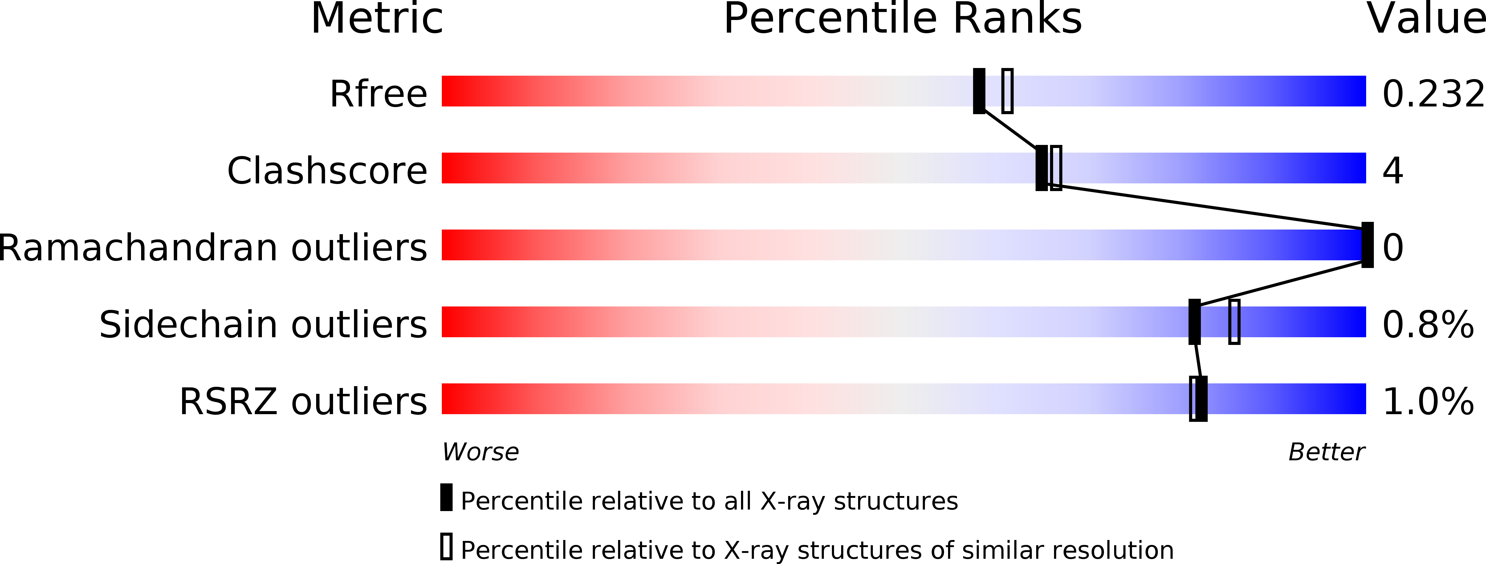
Resolution:
2.00 Å
R-Value Free:
0.22
R-Value Work:
0.17
R-Value Observed:
0.17
Space Group:
P 1 21 1
Macromolecular Entities
Structures with similar UniProt ID
Protein Blast
Polymer Type:polypeptide(L)
Molecule:Uncharacterized protein YgjK
Gene (Uniprot):ygjK
Mutagens:E727A
Chain IDs:A, B
Chain Length:760
Number of Molecules:2
Biological Source:Escherichia coli
Primary Citation
Structure of a bacterial glycoside hydrolase family 63 enzyme in complex with its glycosynthase product, and insights into the substrate specificity.
Febs J. 280 4560 4571 (2013)
PMID: 23826932 DOI: 10.1111/febs.12424

Abstact

Proteins belonging to glycoside hydrolase family 63 (GH63) are found in bacteria, archaea and eukaryotes. Although the eukaryotic GH63 proteins have been identified as processing α-glucosidase I, the substrate specificities of the bacterial and archaeal GH63 proteins are not clear. Here, we converted a bacterial GH63 enzyme, Escherichia coli YgjK, to a glycosynthase to probe its substrate specificity. Two mutants of YgjK (E727A and D324N) were constructed, and both mutants showed glycosynthase activity. The reactions of E727A with β-D-glucosyl fluoride and monosaccharides showed that the largest amount of glycosynthase product accumulated when galactose was employed as an acceptor molecule. The crystal structure of E727A complexed with the reaction product indicated that the disaccharide bound at the active site was 2-O-α-D-glucopyranosyl-α-D-galactopyranose (Glc12Gal). A comparison of the structures of E727A-Glc12Gal and D324N-melibiose showed that there were two main types of conformation: the open and closed forms. The structure of YgjK adopted the closed form when subsite -1 was occupied by glucose. These results suggest that sugars containing the Glc12Gal structure are the most likely candidates for natural substrates of YgjK.

Legend

Protein

Chemical

Disease

Primary Citation of related structures