3W7B image
Deposition Date 2013-02-28
Release Date 2014-01-08
Last Version Date 2023-11-08
Entry Detail
PDB ID:
3W7B
Keywords:
Title:
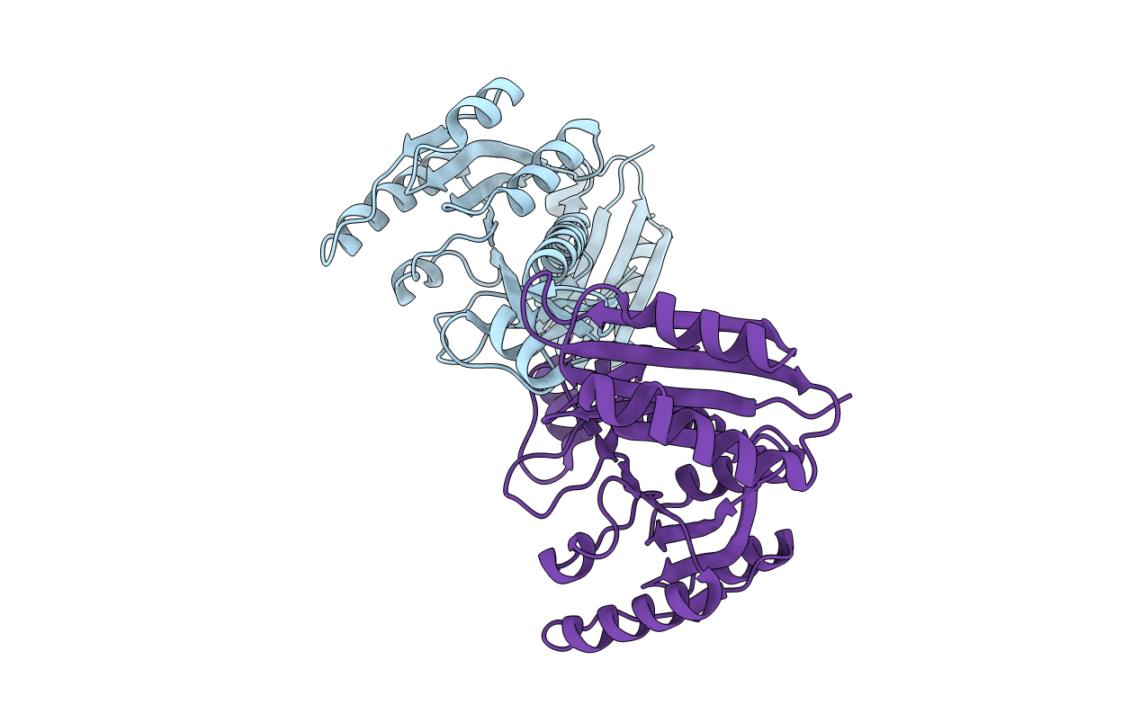
Crystal structure of formyltetrahydrofolate deformylase from Thermus thermophilus HB8
Biological Source:
Source Organism:
Host Organism:
Method Details:
Experimental Method:
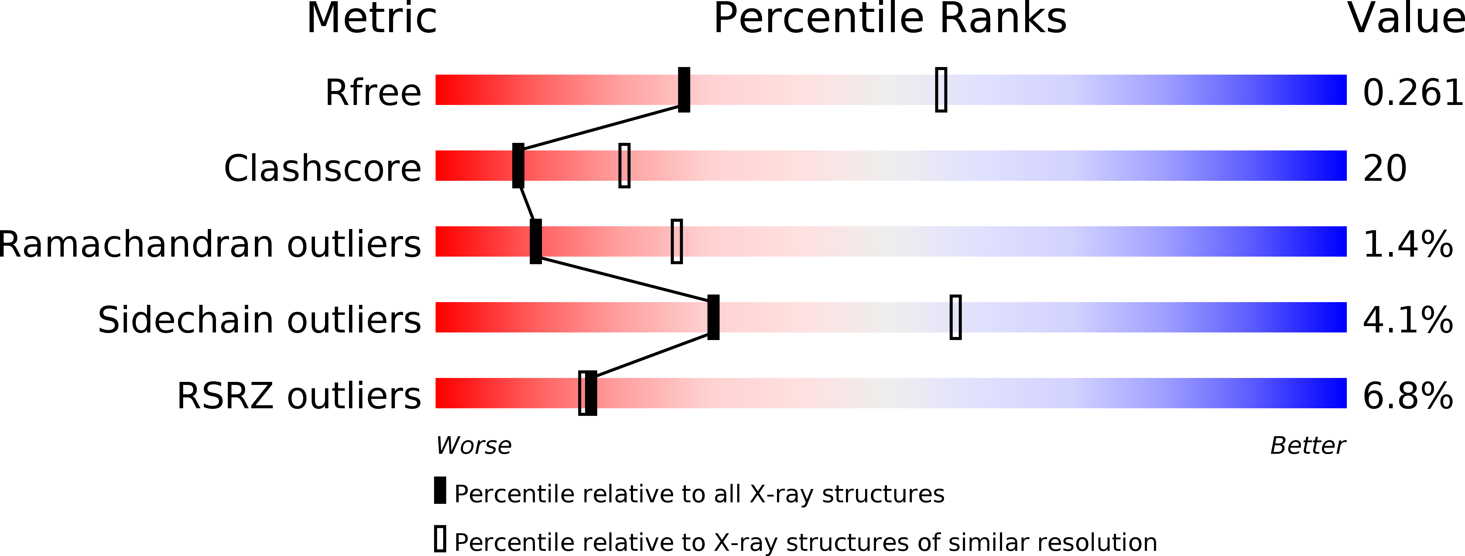
Resolution:
2.71 Å
R-Value Free:
0.26
R-Value Work:
0.20
R-Value Observed:
0.20
Space Group:
P 21 21 2
Macromolecular Entities
Structures with similar UniProt ID
Protein Blast
Polymer Type:polypeptide(L)
Molecule:Formyltetrahydrofolate deformylase
Gene (Uniprot):purU
Chain IDs:A, B
Chain Length:285
Number of Molecules:2
Biological Source:Thermus thermophilus
Primary Citation
Structures and reaction mechanisms of the two related enzymes, PurN and PurU
J.Biochem. 154 569 579 (2013)
PMID: 24108189 DOI: 10.1093/jb/mvt090

Abstact

The crystal structures of glycinamide ribonucleotide transformylases (PurNs) from Aquifex aeolicus (Aa), Geobacillus kaustophilus (Gk) and Symbiobacterium toebii (St), and of formyltetrahydrofolate hydrolase (PurU) from Thermus thermophilus (Tt) were determined. The monomer structures of the determined PurN and PurU were very similar to the known structure of PurN, but oligomeric states were different; AaPurN and StPurN formed dimers, GkPurN formed monomer and PurU formed tetramer in the crystals. PurU had a regulatory ACT domain in its N-terminal side. So far several structures of PurUs have been determined, yet, the mechanisms of the catalysis and the regulation of PurU have not been elucidated. We, therefore, modelled ligand-bound structures of PurN and PurU, and performed molecular dynamics simulations to elucidate the reaction mechanisms. The evolutionary relationship of the two enzymes is discussed based on the comparisons of the structures and the catalytic mechanisms.

Legend

Protein

Chemical

Disease

Primary Citation of related structures
Feedback Form
Name
Email
Institute
Feedback