3W6U image
Deposition Date 2013-02-22
Release Date 2014-01-15
Last Version Date 2024-11-13
Entry Detail
PDB ID:
3W6U
Keywords:
Title:
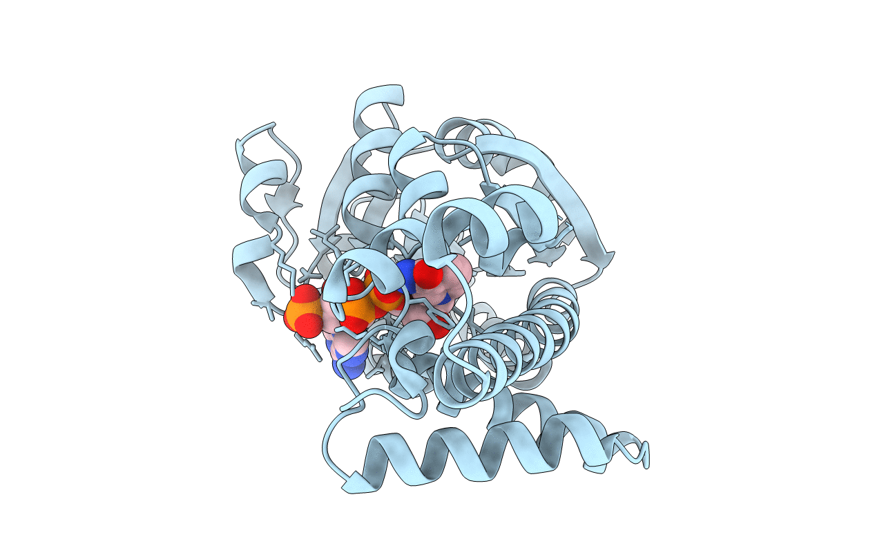
Crystal structure of NADP bound L-serine 3-dehydrogenase from Hyperthermophilic Archaeon Pyrobaculum calidifontis
Biological Source:
Source Organism:
Host Organism:
Method Details:
Experimental Method:
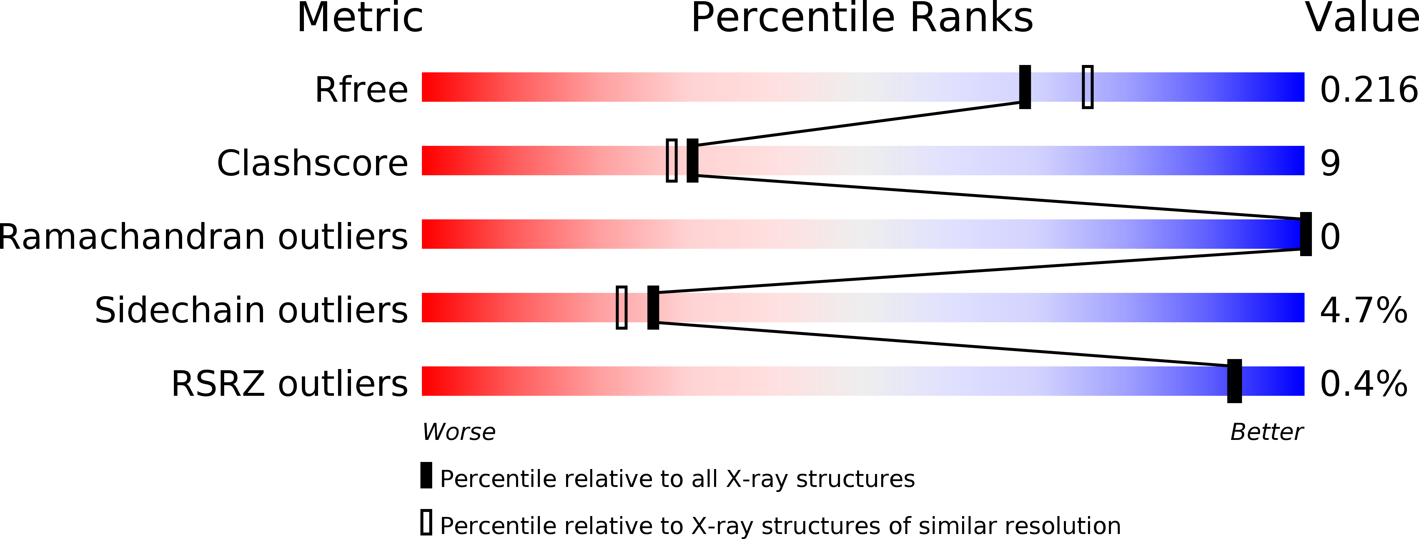
Resolution:
2.00 Å
R-Value Free:
0.24
R-Value Work:
0.22
Space Group:
C 1 2 1
Macromolecular Entities
Polymer Type:polypeptide(L)
Molecule:6-phosphogluconate dehydrogenase, NAD-binding protein
Gene (Uniprot):Pcal_0699
Chain IDs:A
Chain Length:306
Number of Molecules:1
Biological Source:Pyrobaculum calidifontis
Modified Residue
Compound ID Chain ID Parent Comp ID Details 2D Image
MSE A MET SELENOMETHIONINE
Ligand Molecules
Primary Citation
Crystal structure of the NADP+and tartrate-bound complex of L-serine 3-dehydrogenase from the hyperthermophilic archaeon Pyrobaculum calidifontis.
Extremophiles 22 395 405 (2018)
PMID: 29353380 DOI: 10.1007/s00792-018-1004-0

Abstact

A gene encoding L-serine dehydrogenase (L-SerDH) that exhibits extremely low sequence identity to the Agrobacterium tumefaciens L-SerDH was identified in the hyperthermophilic archaeon Pyrobaculum calidifontis. The predicted amino acid sequence showed 36% identity with that of Pseudomonas aeruginosa L-SerDH, suggesting that P. calidifontis L-SerDH is a novel type of L-SerDH, like Ps. aeruginosa L-SerDH. The overexpressed enzyme appears to be the most thermostable L-SerDH described to date, and no loss of activity was observed by incubation for 30 min at temperatures up to 100 °C. The enzyme showed substantial reactivity towards D-serine, in addition to L-serine. Two different crystal structures of P. calidifontis L-SerDH were determined using the Se-MAD and MR method: the structure in complex with NADP+/sulfate ion at 1.18 Å and the structure in complex with NADP+/L-tartrate (substrate analog) at 1.57 Å. The fold of the catalytic domain showed similarity with that of Ps. aeruginosa L-SerDH. However, the active site structure significantly differed between the two enzymes. Based on the structure of the tartrate, L- and D-serine and 3-hydroxypropionate molecules were modeled into the active site and the substrate binding modes were estimated. A structural comparison suggests that the wide cavity at the substrate binding site is likely responsible for the high reactivity of the enzyme toward both L- and D-serine enantiomers. This is the first description of the structure of the novel type of L-SerDH with bound NADP+ and substrate analog, and it provides new insight into the substrate binding mechanism of L-SerDH. The results obtained here may be very informative for the creation of L- or D-serine-specific SerDH by protein engineering.

Legend

Protein

Chemical

Disease

Primary Citation of related structures