Deposition Date
2013-02-06
Release Date
2013-06-26
Last Version Date
2023-11-08
Entry Detail
PDB ID:
3W5T
Keywords:
Title:
Crystal structure of complexes of vitamin D receptor ligand binding domain with lithocholic acid derivatives
Biological Source:
Source Organism(s):
Rattus norvegicus (Taxon ID: 10116)
Homo sapiens (Taxon ID: 9606)
Homo sapiens (Taxon ID: 9606)
Expression System(s):
Method Details:
Experimental Method:
Resolution:
2.29 Å
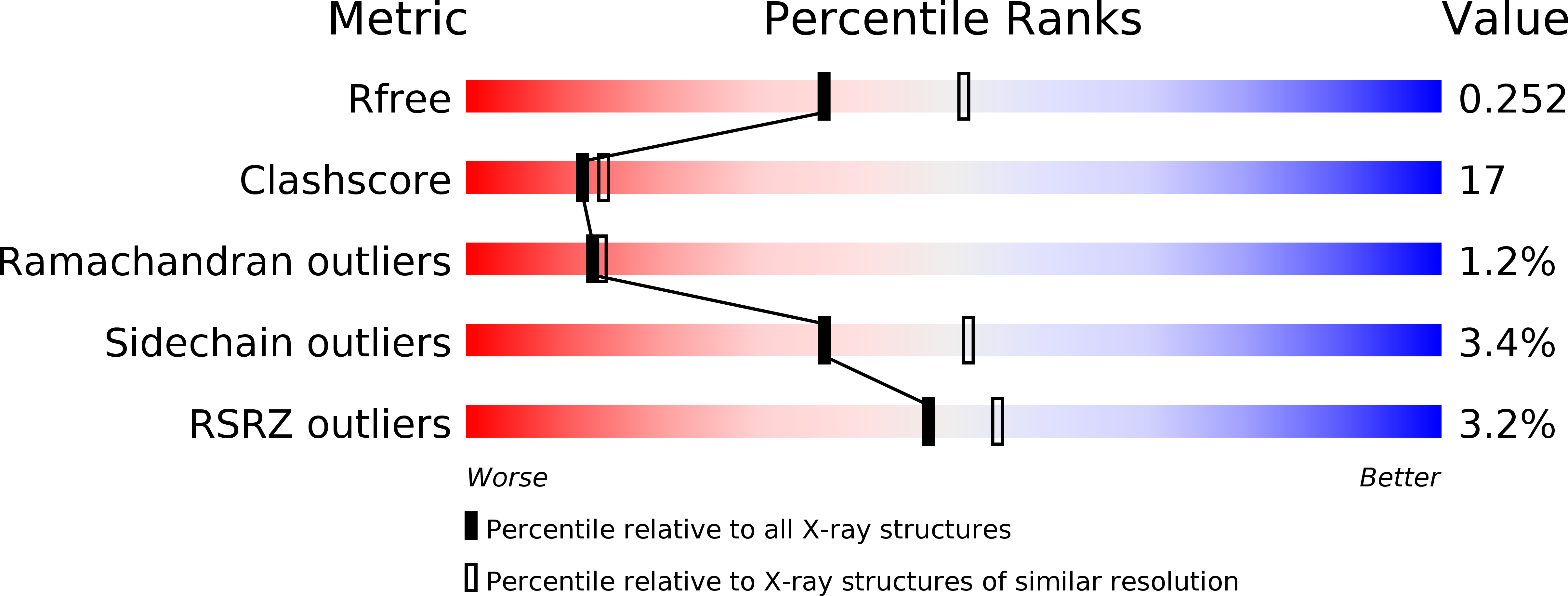
R-Value Free:
0.26
R-Value Work:
0.21
R-Value Observed:
0.21
Space Group:
C 1 2 1