3W5H image
Deposition Date 2013-01-30
Release Date 2013-07-17
Last Version Date 2024-04-03
Entry Detail
PDB ID:
3W5H
Keywords:
Title:
Ultra-high resolution structure of NADH-cytochrome b5 reductase
Biological Source:
Source Organism:
Sus scrofa (Taxon ID: 9823)
Host Organism:
Method Details:
Experimental Method:
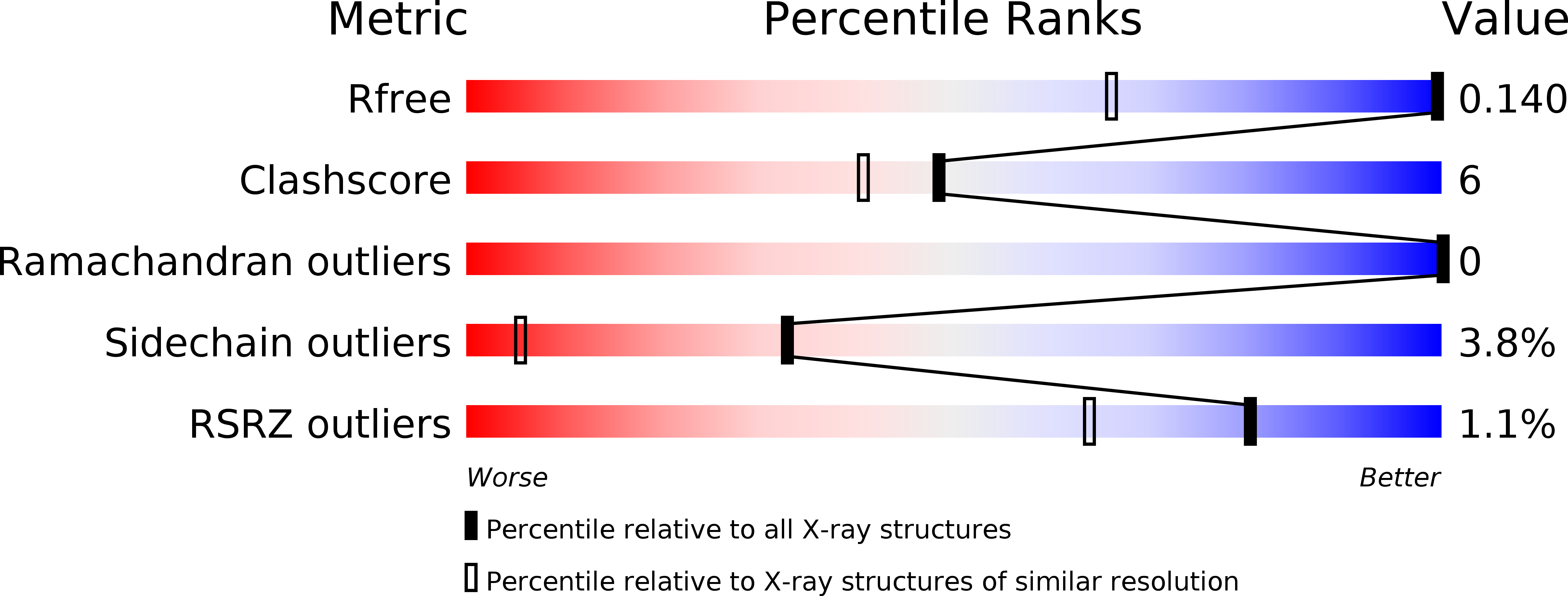
Resolution:
0.78 Å
R-Value Free:
0.14
R-Value Work:
0.12
R-Value Observed:
0.12
Space Group:
P 21 21 21
Macromolecular Entities
Structures with similar UniProt ID
Protein Blast
Polymer Type:polypeptide(L)
Molecule:NADH-cytochrome b5 reductase 3
Gene (Uniprot):CYB5R3
Chain IDs:A
Chain Length:272
Number of Molecules:1
Biological Source:Sus scrofa
Primary Citation
Elucidations of the catalytic cycle of NADH-cytochrome b5 reductase by X-ray crystallography: new insights into regulation of efficient electron transfer
J.Mol.Biol. 425 4295 4306 (2013)
PMID: 23831226 DOI: 10.1016/j.jmb.2013.06.010

Abstact

NADH-Cytochrome b5 reductase (b5R), a flavoprotein consisting of NADH and flavin adenine dinucleotide (FAD) binding domains, catalyzes electron transfer from the two-electron carrier NADH to the one-electron carrier cytochrome b5 (Cb5). The crystal structures of both the fully reduced form and the oxidized form of porcine liver b5R were determined. In the reduced b5R structure determined at 1.68Å resolution, the relative configuration of the two domains was slightly shifted in comparison with that of the oxidized form. This shift resulted in an increase in the solvent-accessible surface area of FAD and created a new hydrogen-bonding interaction between the N5 atom of the isoalloxazine ring of FAD and the hydroxyl oxygen atom of Thr66, which is considered to be a key residue in the release of a proton from the N5 atom. The isoalloxazine ring of FAD in the reduced form is flat as in the oxidized form and stacked together with the nicotinamide ring of NAD(+). Determination of the oxidized b5R structure, including the hydrogen atoms, determined at 0.78Å resolution revealed the details of a hydrogen-bonding network from the N5 atom of FAD to His49 via Thr66. Both of the reduced and oxidized b5R structures explain how backflow in this catalytic cycle is prevented and the transfer of electrons to one-electron acceptors such as Cb5 is accelerated. Furthermore, crystallographic analysis by the cryo-trapping method suggests that re-oxidation follows a two-step mechanism. These results provide structural insights into the catalytic cycle of b5R.

Legend

Protein

Chemical

Disease

Primary Citation of related structures