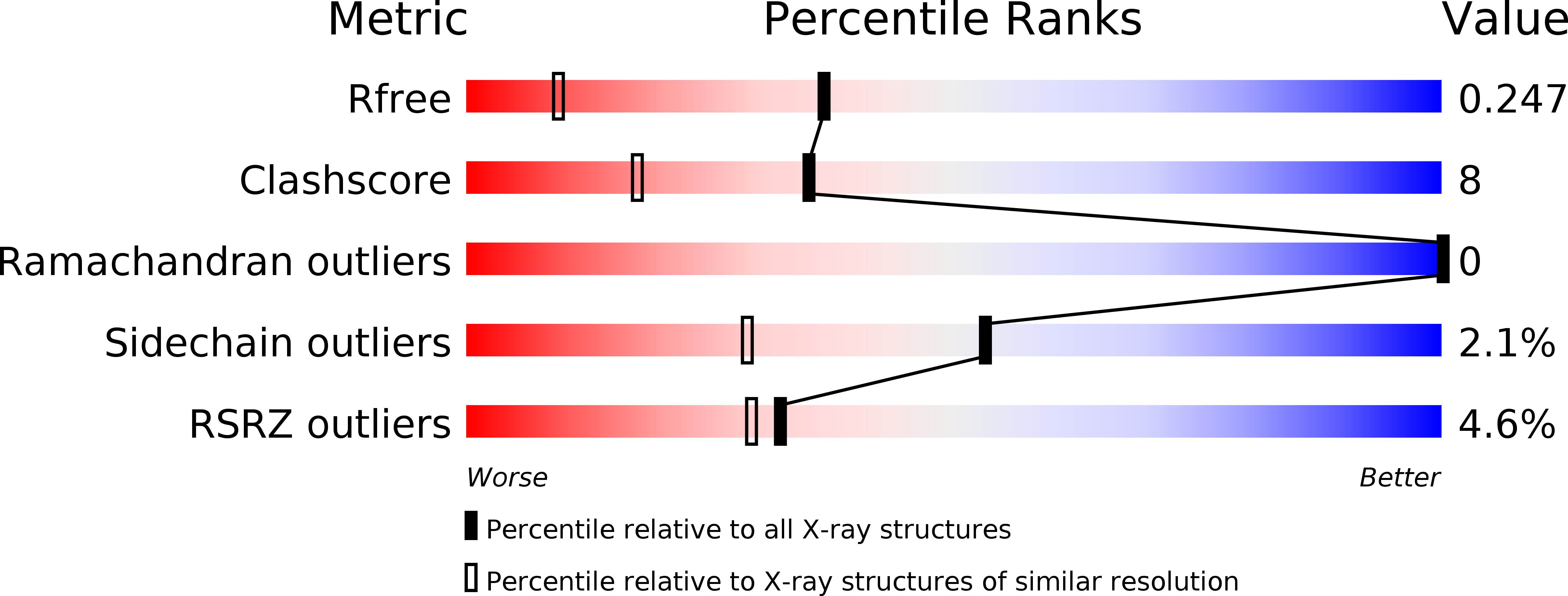
3W56 image
Entry Detail
PDB ID:
3W56
Title:
Structure of a C2 domain
Biological Source:
Source Organism:
Host Organism:
PDB Version:
Deposition Date:
2013-01-24
Release Date:
2013-10-23
Method Details:
Experimental Method:
Resolution:
1.60 Å
R-Value Free:
0.25
R-Value Work:
0.21
R-Value Observed:
0.21
Space Group:
I 1 2 1
Macromolecular Entities
Polymer Type:polypeptide(L)
Description:C2 domain protein
Chain IDs:A
Chain Length:131
Number of Molecules:1
Biological Source:Scophthalmus maximus
Primary Citation
Defining the interaction of perforin with calcium and the phospholipid membrane.
Biochem.J. 456 323 335 (2013)
PMID: 24070258 DOI: 10.1042/BJ20130999

Abstact

Following its secretion from cytotoxic lymphocytes into the immune synapse, perforin binds to target cell membranes through its Ca(2+)-dependent C2 domain. Membrane-bound perforin then forms pores that allow passage of pro-apoptopic granzymes into the target cell. In the present study, structural and biochemical studies reveal that Ca(2+) binding triggers a conformational change in the C2 domain that permits four key hydrophobic residues to interact with the plasma membrane. However, in contrast with previous suggestions, these movements and membrane binding do not trigger irreversible conformational changes in the pore-forming MACPF (membrane attack complex/perforin-like) domain, indicating that subsequent monomer-monomer interactions at the membrane surface are required for perforin pore formation.

Legend

Protein

Chemical

Disease

Primary Citation of related structures