Deposition Date
2012-11-27
Release Date
2013-09-04
Last Version Date
2024-10-09
Entry Detail
PDB ID:
3W2A
Keywords:
Title:
Crystal structure of VirB core domain complexed with the cis-acting site upstream icsp promoter
Biological Source:
Source Organism(s):
Shigella flexneri 2a (Taxon ID: 42897)
Expression System(s):
Method Details:
Experimental Method:
Resolution:
2.78 Å
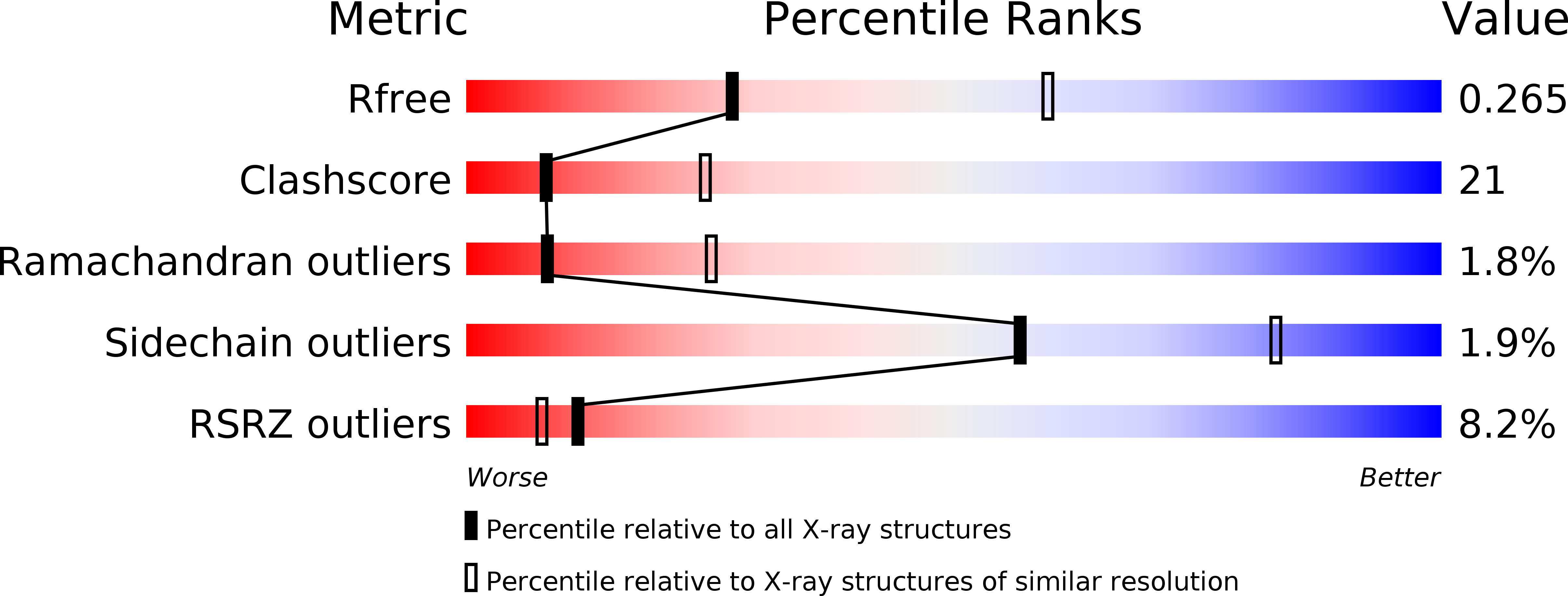
R-Value Free:
0.26
R-Value Work:
0.24
R-Value Observed:
0.24
Space Group:
C 1 2 1