Deposition Date
2012-10-15
Release Date
2013-08-28
Last Version Date
2023-11-08
Entry Detail
PDB ID:
3VZS
Keywords:
Title:
Crystal structure of PhaB from Ralstonia eutropha in complex with Acetoacetyl-CoA and NADP
Biological Source:
Source Organism(s):
Cupriavidus necator (Taxon ID: 381666)
Expression System(s):
Method Details:
Experimental Method:
Resolution:
2.14 Å
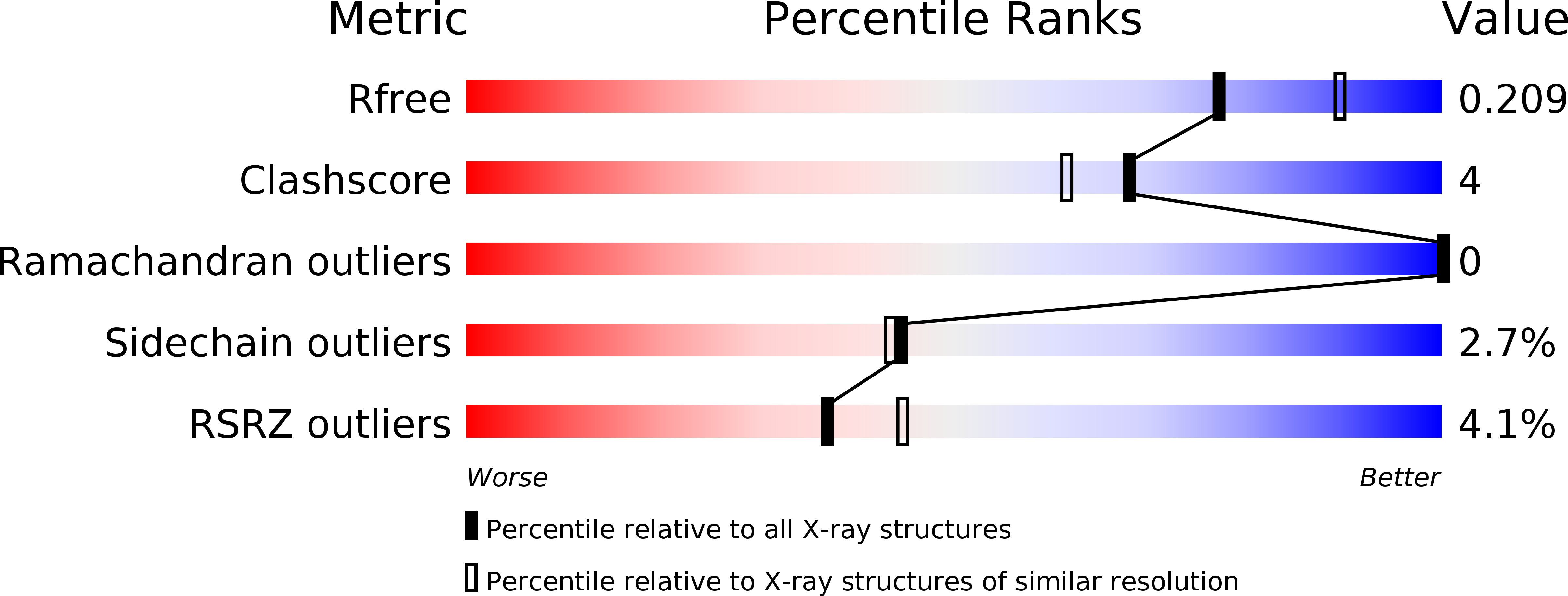
R-Value Free:
0.20
R-Value Work:
0.16
R-Value Observed:
0.16
Space Group:
C 2 2 21