3VYU image
Deposition Date 2012-10-02
Release Date 2012-11-28
Last Version Date 2024-10-16
Entry Detail
PDB ID:
3VYU
Title:
Crystal structure of the HypC-HypD-HypE complex (form II)
Biological Source:
Source Organism:
Host Organism:
Method Details:
Experimental Method:
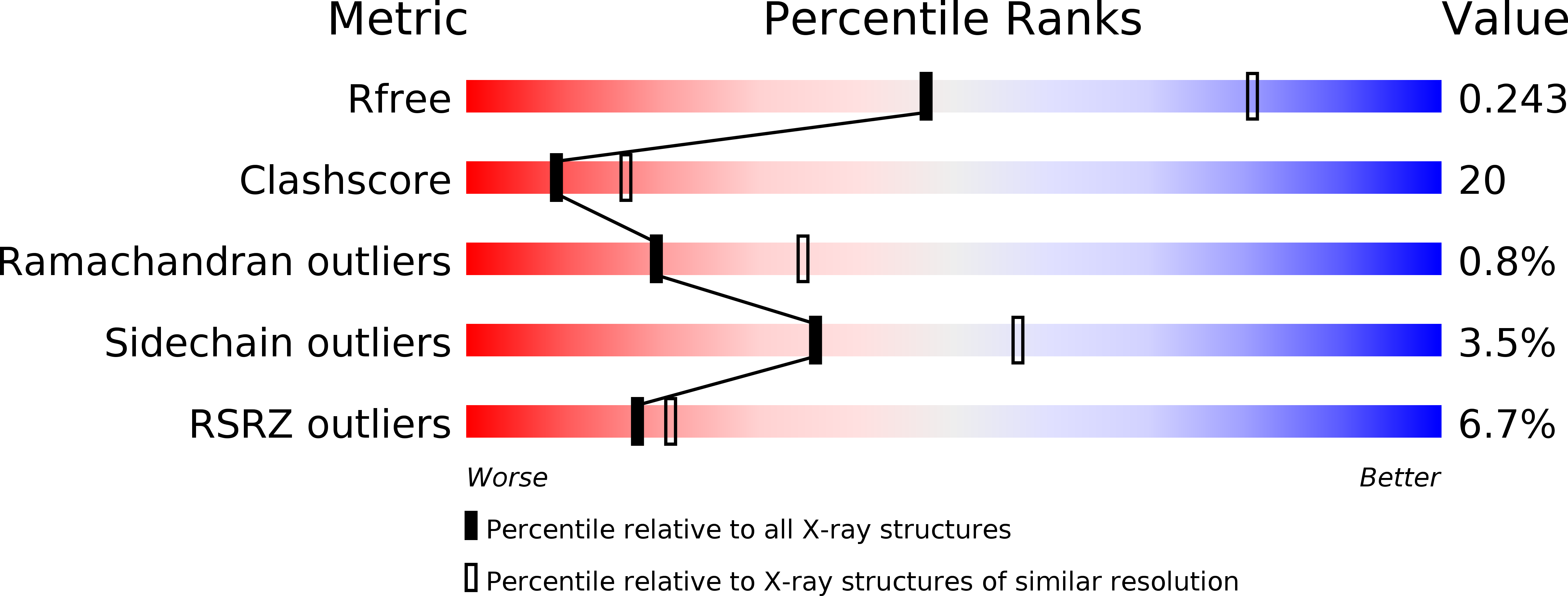
Resolution:
2.75 Å
R-Value Free:
0.25
R-Value Work:
0.21
R-Value Observed:
0.21
Space Group:
C 1 2 1
Macromolecular Entities
Structures with similar UniProt ID
Protein Blast
Polymer Type:polypeptide(L)
Molecule:Hydrogenase expression/formation protein HypC
Gene (Uniprot):Tk-hypC
Chain IDs:A
Chain Length:74
Number of Molecules:1
Biological Source:Thermococcus kodakarensis
Structures with similar UniProt ID
Protein Blast
Polymer Type:polypeptide(L)
Molecule:Hydrogenase expression/formation protein HypD
Gene (Uniprot):Tk-hypD
Chain IDs:B
Chain Length:372
Number of Molecules:1
Biological Source:Thermococcus kodakarensis
Structures with similar UniProt ID
Protein Blast
Polymer Type:polypeptide(L)
Molecule:Hydrogenase expression/formation protein HypE
Gene (Uniprot):TK1993
Chain IDs:C
Chain Length:338
Number of Molecules:1
Biological Source:Thermococcus kodakarensis
Ligand Molecules
Primary Citation
Crystal structures of the HypCD complex and the HypCDE ternary complex: transient intermediate complexes during [NiFe] hydrogenase maturation
Structure 20 2124 2137 (2012)
PMID: 23123111 DOI: 10.1016/j.str.2012.09.018

Abstact

[NiFe] hydrogenase maturation represents one of the most dynamic and sophisticated processes in metallocenter assembly. The Fe(CN)(2)CO moiety of [NiFe] hydrogenases is assembled via unknown transient interactions among specific maturation proteins HypC (metallochaperone), HypD (redox protein), and HypE (cyanide synthesis/donor). Here, we report the structures of the HypC-HypD and HypC-HypD-HypE complexes, providing a view of the transient interactions that take place during the maturation process. HypC binds to the conserved region of HypD through extensive hydrophobic interactions. The ternary complex formation between HypE and the HypCD complex involves both HypC and HypD, rendering the HypE conformation favorable for cyanide transfer. In the complex, the conserved cysteines of HypC and HypD form an Fe binding site. The conserved C-terminal cysteine of HypE can access the thiol redox cascade of HypD. These results provide structural insights into the Fe atom cyanation in the HypCDE complex.

Legend

Protein

Chemical

Disease

Primary Citation of related structures
Feedback Form
Name
Email
Institute
Feedback