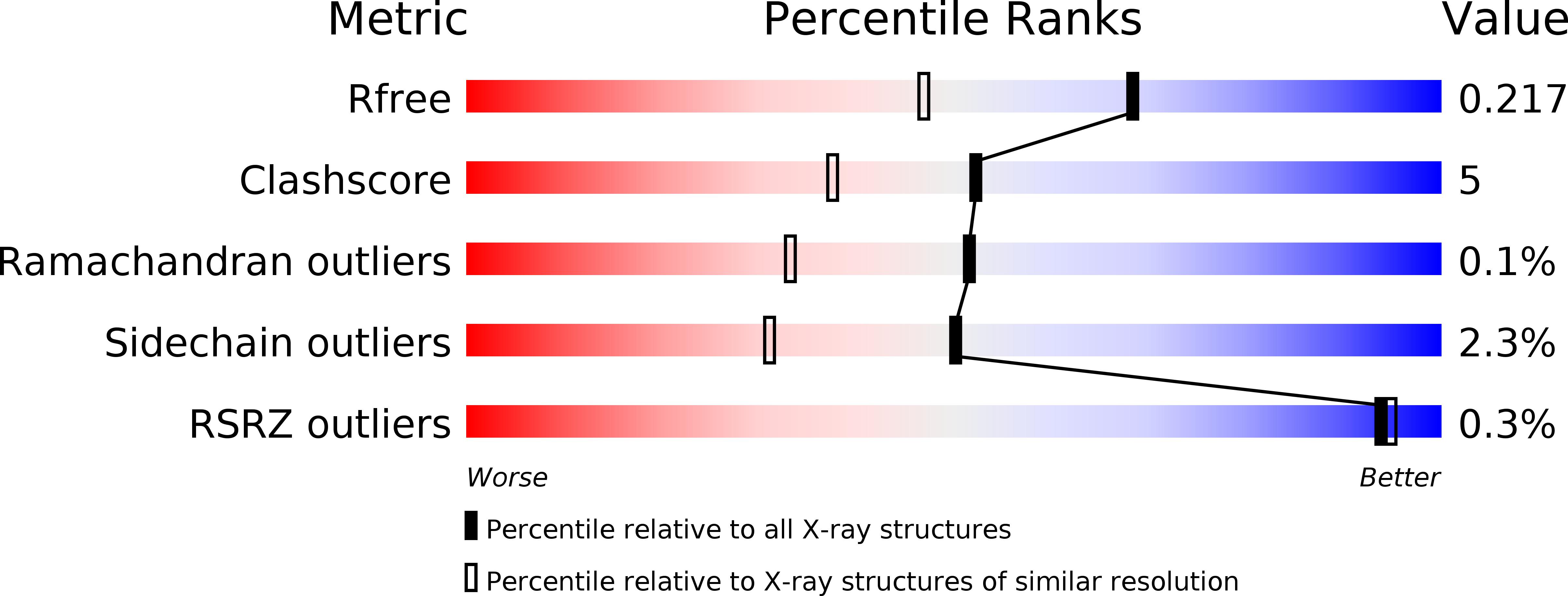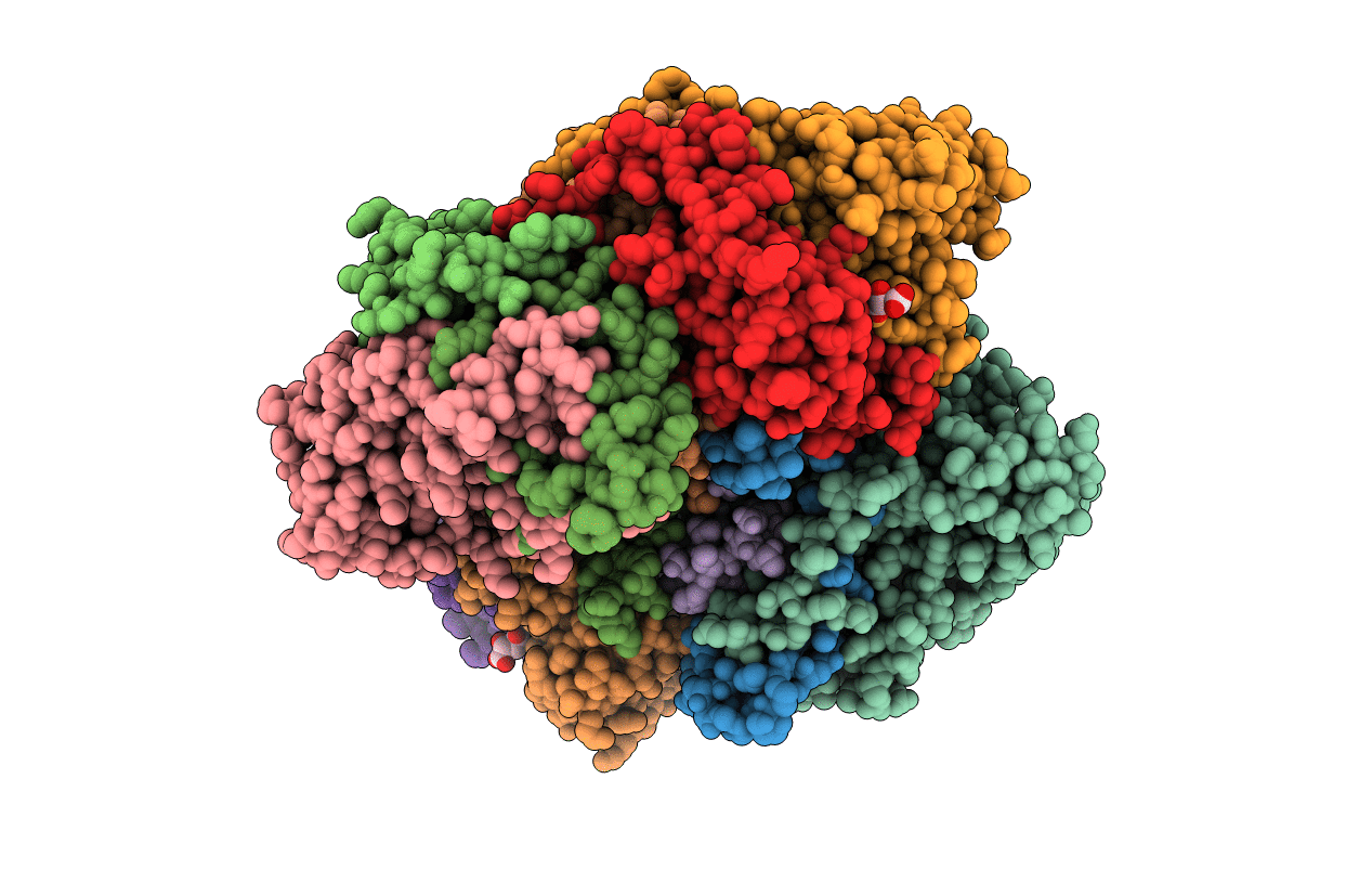
3VYG image
Entry Detail
PDB ID:
3VYG
Keywords:
Title:
Crystal structure of Thiocyanate hydrolase mutant R136W
Biological Source:
Source Organism:
Host Organism:
PDB Version:
Deposition Date:
2012-09-25
Release Date:
2013-11-13
Method Details:
Experimental Method:
Resolution:
1.72 Å
R-Value Free:
0.21
R-Value Work:
0.18
R-Value Observed:
0.18
Space Group:
P 21 21 21
Macromolecular Entities
Polymer Type:polypeptide(L)
Description:Thiocyanate hydrolase subunit alpha
Chain IDs:A, D, G, J
Chain Length:126
Number of Molecules:4
Biological Source:Thiobacillus thioparus
Polymer Type:polypeptide(L)
Description:Thiocyanate hydrolase subunit beta
Chain IDs:B, E, H, K
Chain Length:157
Number of Molecules:4
Biological Source:Thiobacillus thioparus
Polymer Type:polypeptide(L)
Description:Thiocyanate hydrolase subunit gamma
Mutations:R136W
Chain IDs:C, F, I, L
Chain Length:243
Number of Molecules:4
Biological Source:Thiobacillus thioparus
Modified Residue
Compound ID Chain ID Parent Comp ID Details 2D Image
CSD C CYS 3-SULFINOALANINE
CSO C CYS S-HYDROXYCYSTEINE
Primary Citation
Effects of argnine residue around the substrate pocket on the substrate specificity of thiocyanate hydrolase
To be published ? ? ? (?)
Primary Citation of related structures