
Deposition Date
2012-07-30
Release Date
2013-03-27
Last Version Date
2024-11-06
Entry Detail
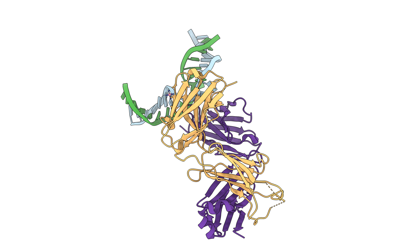
PDB ID:
3VW3
Keywords:
Title:
Antibody 64M-5 Fab in complex with a double-stranded DNA (6-4) photoproduct
Biological Source:
Source Organism(s):
Mus musculus (Taxon ID: 10090)
Method Details:
Experimental Method:
Resolution:
2.50 Å
R-Value Free:
0.29
R-Value Work:
0.24
R-Value Observed:
0.24
Space Group:
P 43 21 2
