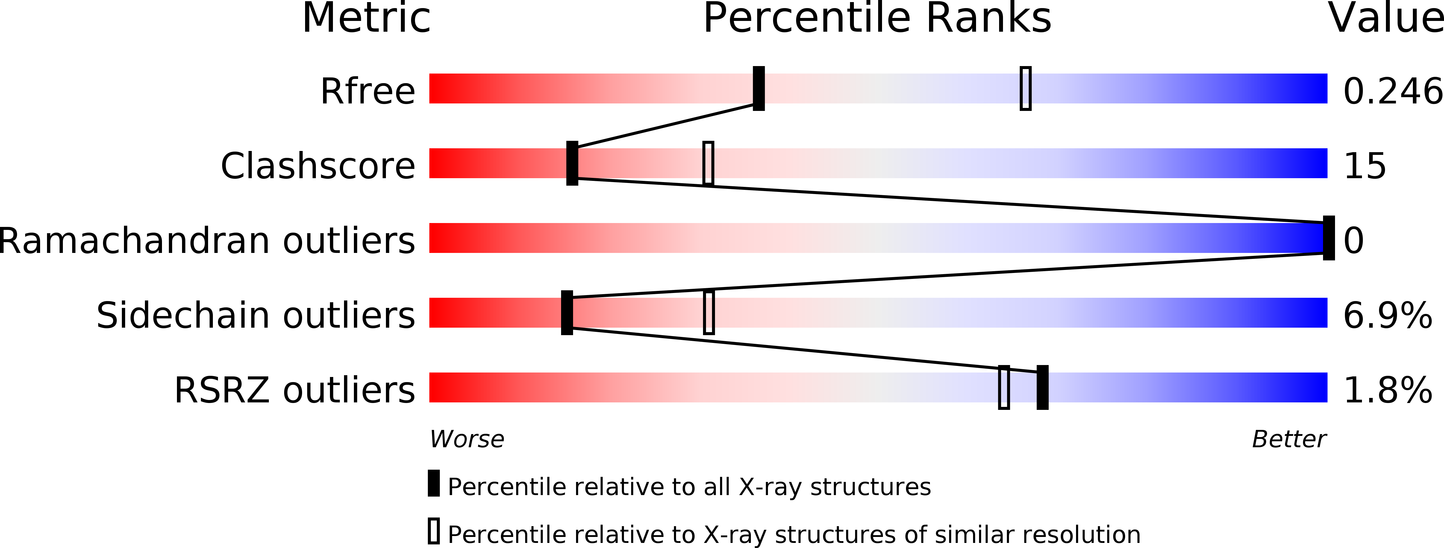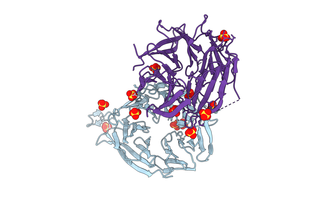
3VU4 image
Deposition Date 2012-06-15
Release Date 2012-07-04
Last Version Date 2024-03-20
Entry Detail
PDB ID:
3VU4
Title:
Crystal structure of Kluyvelomyces marxianus Hsv2
Biological Source:
Source Organism:
Host Organism:
Method Details:
Experimental Method:
Resolution:
2.60 Å
R-Value Free:
0.25
R-Value Work:
0.22
R-Value Observed:
0.22
Space Group:
P 65 2 2
Macromolecular Entities
Polymer Type:polypeptide(L)
Molecule:KmHsv2
Chain IDs:A, B
Chain Length:355
Number of Molecules:2
Biological Source:Kluyveromyces marxianus
Ligand Molecules
Primary Citation
Structure-based analyses reveal distinct binding sites for Atg2 and phosphoinositides in Atg18.
J.Biol.Chem. 287 31681 31690 (2012)
PMID: 22851171 DOI: 10.1074/jbc.M112.397570

Abstact

Autophagy is an intracellular degradation system by which cytoplasmic materials are enclosed by an autophagosome and delivered to a lysosome/vacuole. Atg18 plays a critical role in autophagosome formation as a complex with Atg2 and phosphatidylinositol 3-phosphate (PtdIns(3)P). However, little is known about the structure of Atg18 and its recognition mode of Atg2 or PtdIns(3)P. Here, we report the crystal structure of Kluyveromyces marxianus Hsv2, an Atg18 paralog, at 2.6 Å resolution. The structure reveals a seven-bladed β-propeller without circular permutation. Mutational analyses of Atg18 based on the K. marxianus Hsv2 structure suggested that Atg18 has two phosphoinositide-binding sites at blades 5 and 6, whereas the Atg2-binding region is located at blade 2. Point mutations in the loops of blade 2 specifically abrogated autophagy without affecting another Atg18 function, the regulation of vacuolar morphology at the vacuolar membrane. This architecture enables Atg18 to form a complex with Atg2 and PtdIns(3)P in parallel, thereby functioning in the formation of autophagosomes at autophagic membranes.

Legend

Protein

Chemical

Disease

Primary Citation of related structures