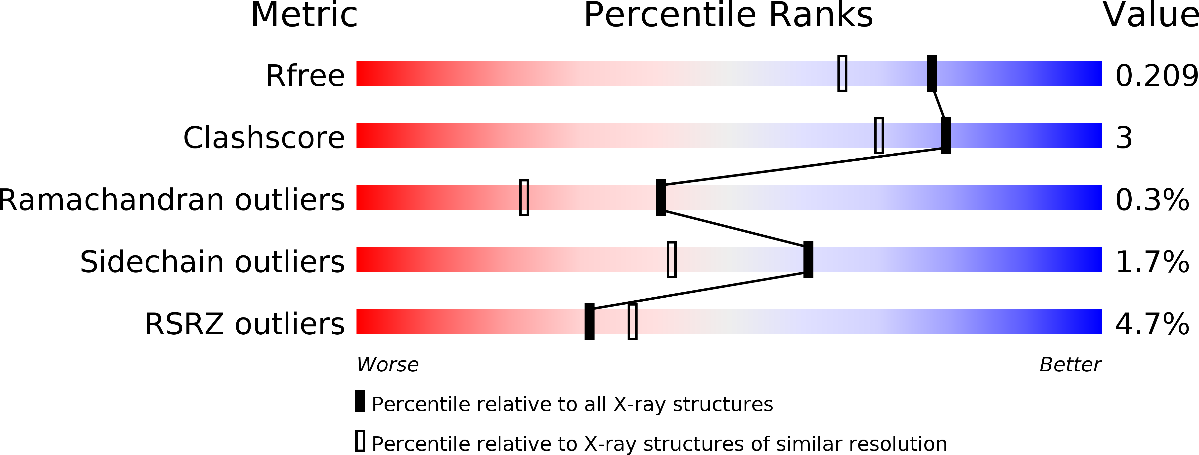
3VTX image
Deposition Date 2012-06-08
Release Date 2012-10-31
Last Version Date 2024-03-20
Entry Detail
PDB ID:
3VTX
Keywords:
Title:
Crystal structure of MamA protein
Biological Source:
Source Organism:
Host Organism:
Method Details:
Experimental Method:
Resolution:
1.75 Å
R-Value Free:
0.20
R-Value Work:
0.16
R-Value Observed:
0.16
Space Group:
P 21 21 21
Macromolecular Entities
Structures with similar UniProt ID
Protein Blast
Polymer Type:polypeptide(L)
Molecule:MamA
Chain IDs:A, B
Chain Length:184
Number of Molecules:2
Biological Source:Candidatus Magnetobacterium bavaricum
Ligand Molecules
Primary Citation
Inter-phylum structural conservation of the magnetosome-associated TPR-containing protein, MamA
J.Struct.Biol. 180 479 487 (2012)
PMID: 22917855 DOI: 10.1016/j.jsb.2012.08.001

Abstact

Magnetotactic bacteria enclose the magnetosome, a unique prokaryotic sub-cellular organelle that allows the biomineralization of magnetic nano-crystals. Membrane-coated magnetosomes are arranged into a linear chain that permits magnetotactic bacteria to navigate geomagnetic fields. Magnetosome assembly and biomineralization are controlled by conserved magnetosome-associated proteins, including MamA, a tetra-trico-peptide repeat (TPR)-containing protein that was shown to coat the magnetosome membrane. In this study, two MamA structures from Candidatus Magnetobacterium bavaricum (Mbav) were determined via X-ray crystallography. These structures confirm that Mbav MamA folds as a sequential TPR protein and shares a high degree of structural similarity with homologous MamA proteins from Magnetospirillum species. Furthermore, the two TPR-containing domains of MamA are separated by an interphylum-conserved region containing a flexible hinge that is involved in ligand binding and recognition. Finally, substantial differences were found in the local stabilization of the MamA N-terminal domain as a result of the loss of an evolutionary conserved salt bridge.

Legend

Protein

Chemical

Disease

Primary Citation of related structures