3VTF image
Deposition Date 2012-05-29
Release Date 2012-09-19
Last Version Date 2023-11-08
Entry Detail
PDB ID:
3VTF
Keywords:
Title:
Structure of a UDP-glucose dehydrogenase from the hyperthermophilic archaeon Pyrobaculum islandicum
Biological Source:
Source Organism:
Host Organism:
Method Details:
Experimental Method:
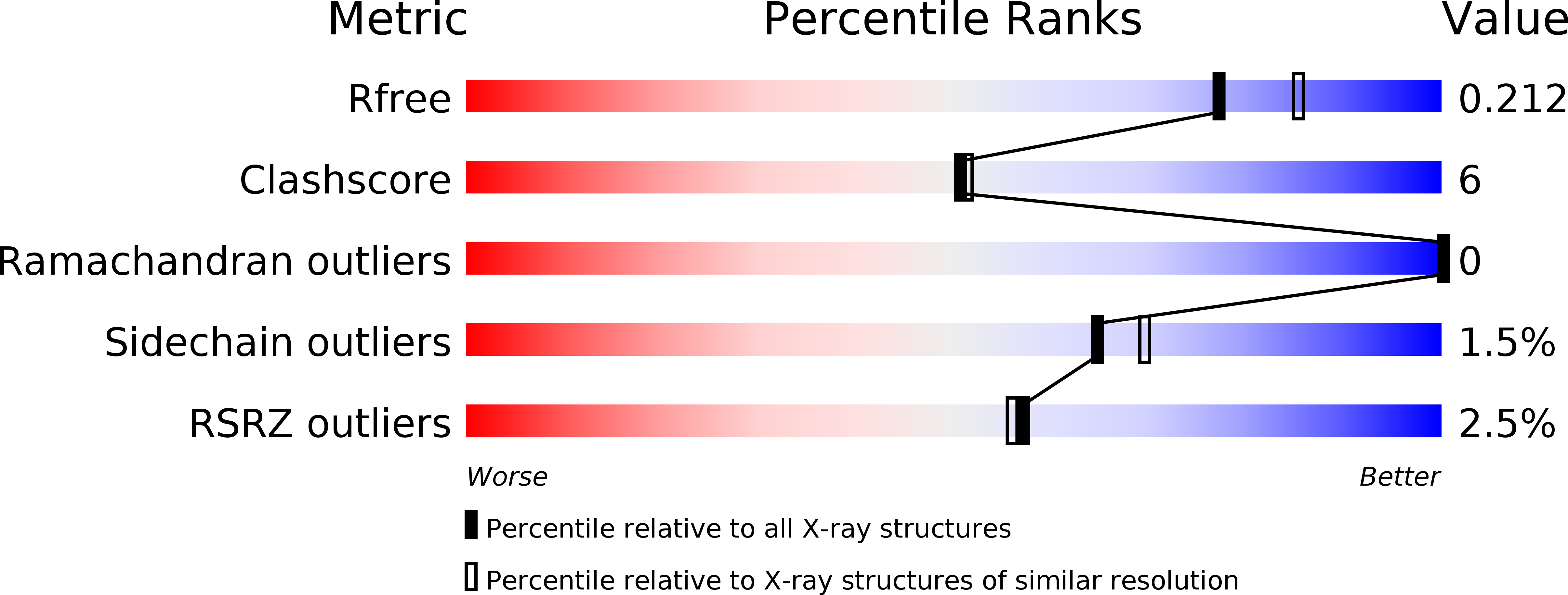
Resolution:
2.00 Å
R-Value Free:
0.21
R-Value Work:
0.17
R-Value Observed:
0.17
Space Group:
C 1 2 1
Macromolecular Entities
Structures with similar UniProt ID
Protein Blast
Polymer Type:polypeptide(L)
Molecule:UDP-glucose 6-dehydrogenase
Gene (Uniprot):Pisl_1505
Chain IDs:A
Chain Length:444
Number of Molecules:1
Biological Source:Pyrobaculum islandicum
Primary Citation
Structure of a UDP-glucose dehydrogenase from the hyperthermophilic archaeon Pyrobaculum islandicum.
Acta Crystallogr.,Sect.F 68 1003 1007 (2012)
PMID: 22949183 DOI: 10.1107/S1744309112030667

Abstact

The crystal structure of an extremely thermostable UDP-glucose dehydrogenase (UDP-GDH) from the hyperthermophilic archaeon Pyrobaculum islandicum was determined at a resolution of 2.0 Å. The overall fold was comprised of an N-terminal NAD(+) dinucleotide binding domain and a C-terminal UDP-sugar binding domain connected by a long α-helix, and the main-chain coordinates of the enzyme were similar to those of previously studied UDP-GDHs, including the enzymes from Burkholderia cepacia, Streptococcus pyogenes and Klebsiella pneumoniae. However, the sizes of several surface loops in P. islandicum UDP-GDH were much smaller than the corresponding loops in B. cepacia UDP-GDH but were comparable to those of the S. pyogenes and K. pneumoniae enzymes. Structural comparison revealed that the presence of extensive intersubunit hydrophobic interactions, as well as the formation of an intersubunit aromatic pair network, is likely to be the main factor contributing to the hyperthermostability of P. islandicum UDP-GDH.

Legend

Protein

Chemical

Disease

Primary Citation of related structures