
Deposition Date
2012-05-19
Release Date
2013-05-22
Last Version Date
2023-11-08
Entry Detail
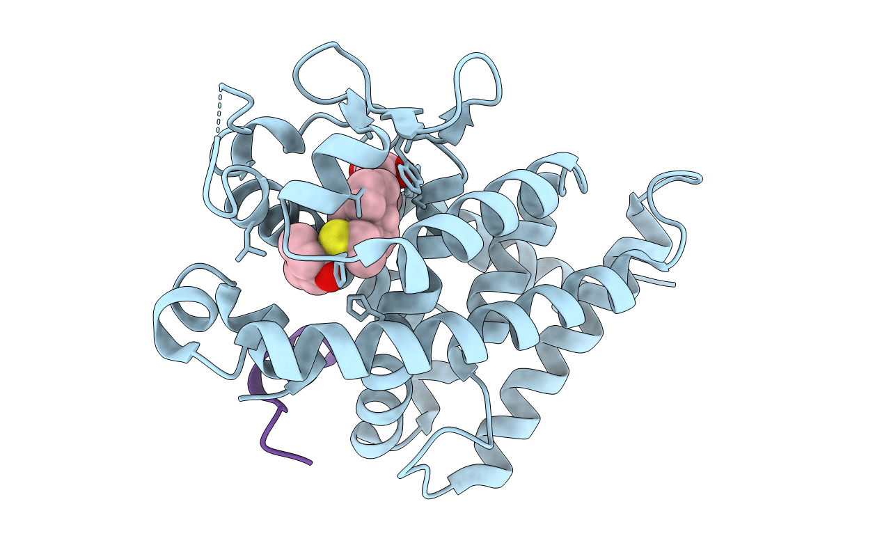
PDB ID:
3VT4
Keywords:
Title:
Crystal structures of rat VDR-LBD with R270L mutation
Biological Source:
Source Organism(s):
Rattus norvegicus (Taxon ID: 10116)
Expression System(s):
Method Details:
Experimental Method:
Resolution:
1.90 Å
R-Value Free:
0.26
R-Value Work:
0.21
R-Value Observed:
0.21
Space Group:
C 1 2 1
