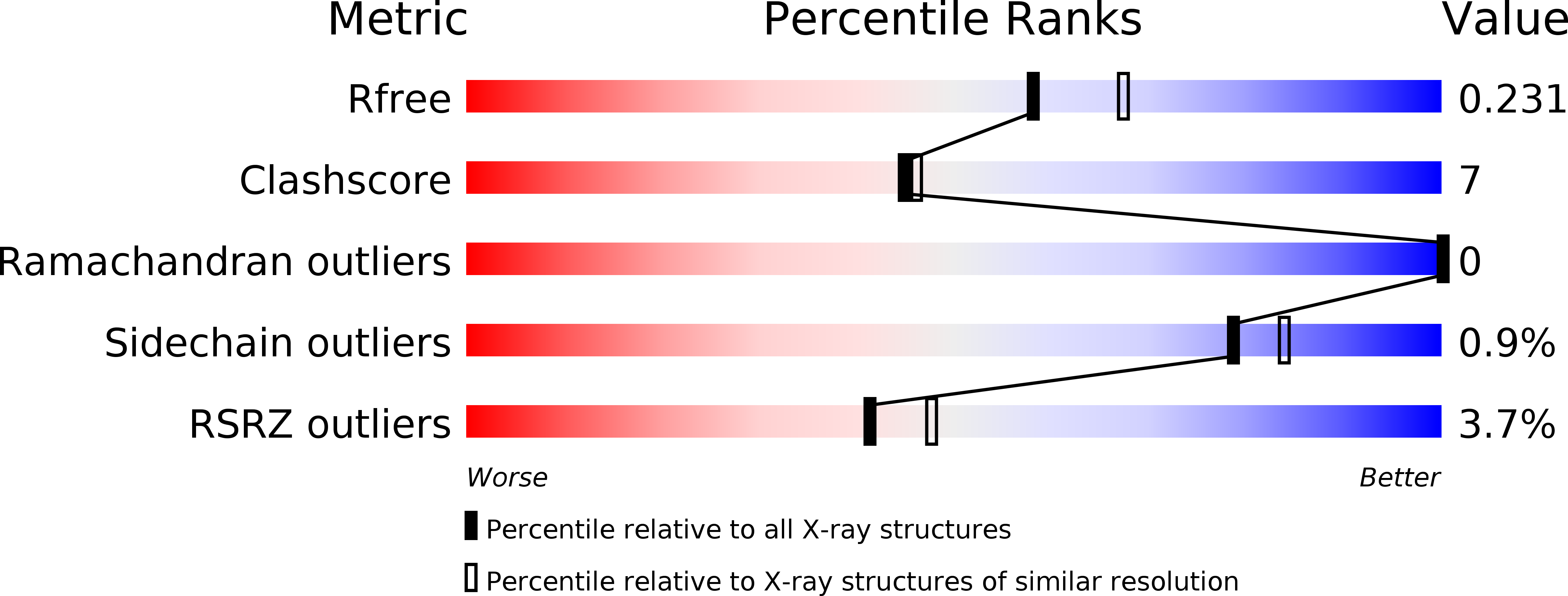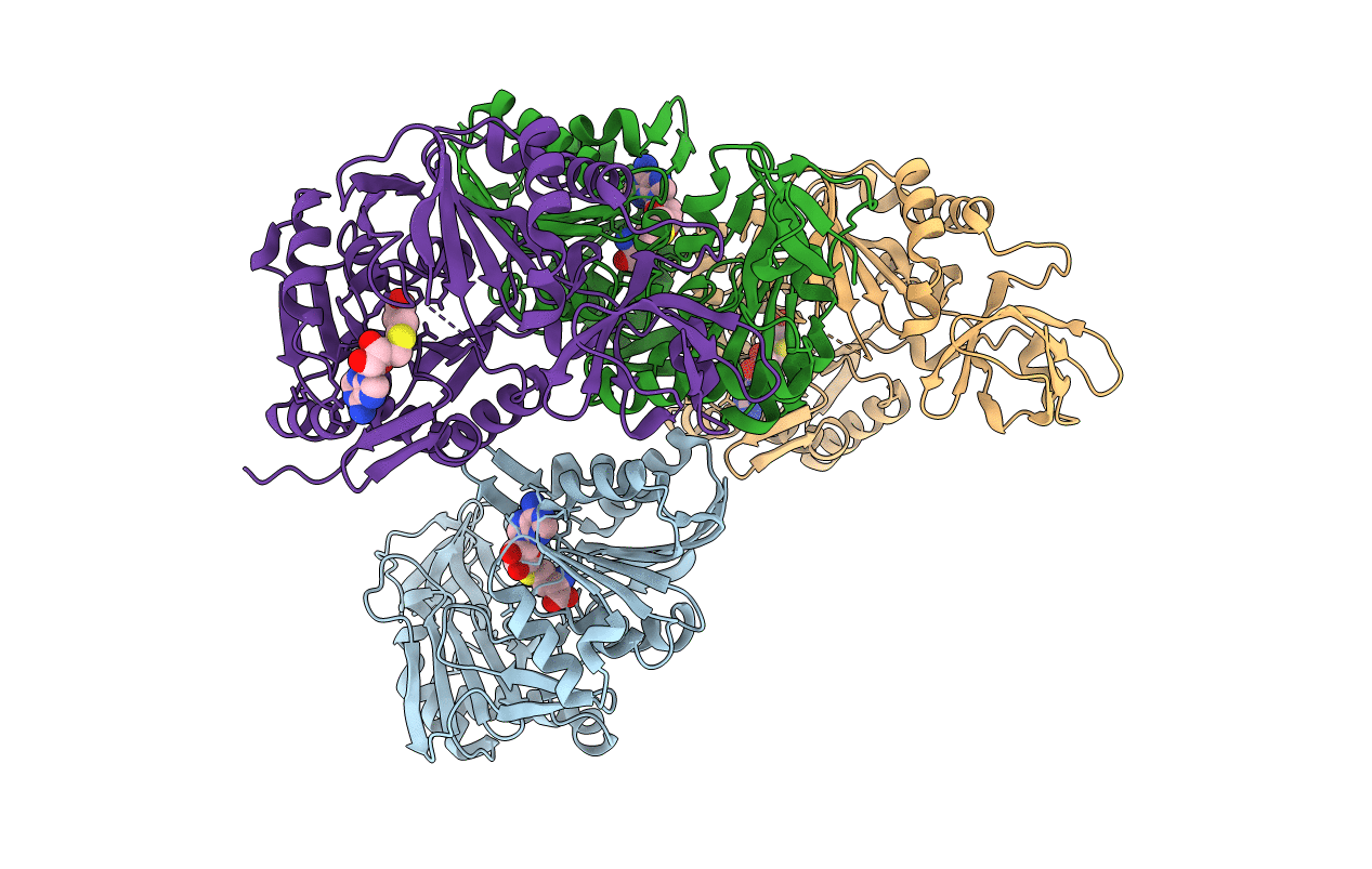
3VSE image
Deposition Date 2012-04-25
Release Date 2013-04-10
Last Version Date 2024-03-20
Entry Detail
PDB ID:
3VSE
Keywords:
Title:
Crystal structure of methyltransferase
Biological Source:
Source Organism:
Host Organism:
Method Details:
Experimental Method:
Resolution:
2.10 Å
R-Value Free:
0.22
R-Value Work:
0.20
R-Value Observed:
0.20
Space Group:
P 1 21 1
Macromolecular Entities
Polymer Type:polypeptide(L)
Molecule:Putative uncharacterized protein
Chain IDs:A, B, C, D
Chain Length:398
Number of Molecules:4
Biological Source:Staphylococcus aureus
Ligand Molecules
Primary Citation
Crystal structure of a putative methyltransferase SAV1081 from Staphylococcus aureus
Protein Pept.Lett. 20 530 537 (2012)
PMID: 23016631 DOI: 10.2174/0929866511320050006

Abstact

Cluster of Orthologous Groups (COG) 1092 contains two distinct types of methylation enzyme from Escherichia coli, YccW and YcbY. YccW is a 5-methylcytosine methyltransferase (m5C MTase) responsible for m5C 1962 in 23S rRNA, whereas YcbY is a dimethyltransferase, of which N- and C-terminal domains are responsible for N2- methylguanosine (m2G) 2445 and 7-methylguanosine (m7G) 2069 in 23S rRNA, respectively. However, proteins in COG1092 other than YccW and YcbY remain functionally unidentified. SAV1081 from Staphylococcus aureus is one of the functionally unassigned proteins of COG1092. Although SAV1081 has an identical domain organization to YccW with 26% sequence identity, it lacks the catalytic cysteine residue essential for m5C formation activity. In the present study, we determined the crystal structure of SAV1081 and compared it with those of other COG1092 proteins. Based on the structure characteristics, such as the presence or absence of the catalytic cysteine residue, β-hairpin structure, and oligomeric state, as well as domain organization, we propose a functional classification of COG1092 proteins.

Legend

Protein

Chemical

Disease

Primary Citation of related structures