3VRD image
Entry Detail
PDB ID:
3VRD
Title:
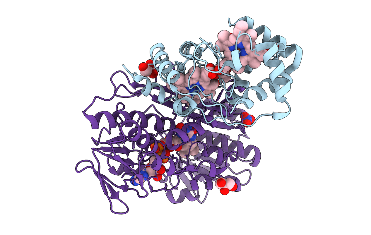
Crystal structure of flavocytochrome c from Thermochromatium tepidum
Biological Source:
Source Organism:
PDB Version:
Deposition Date:
2012-04-09
Release Date:
2012-09-12
Method Details:
Experimental Method:
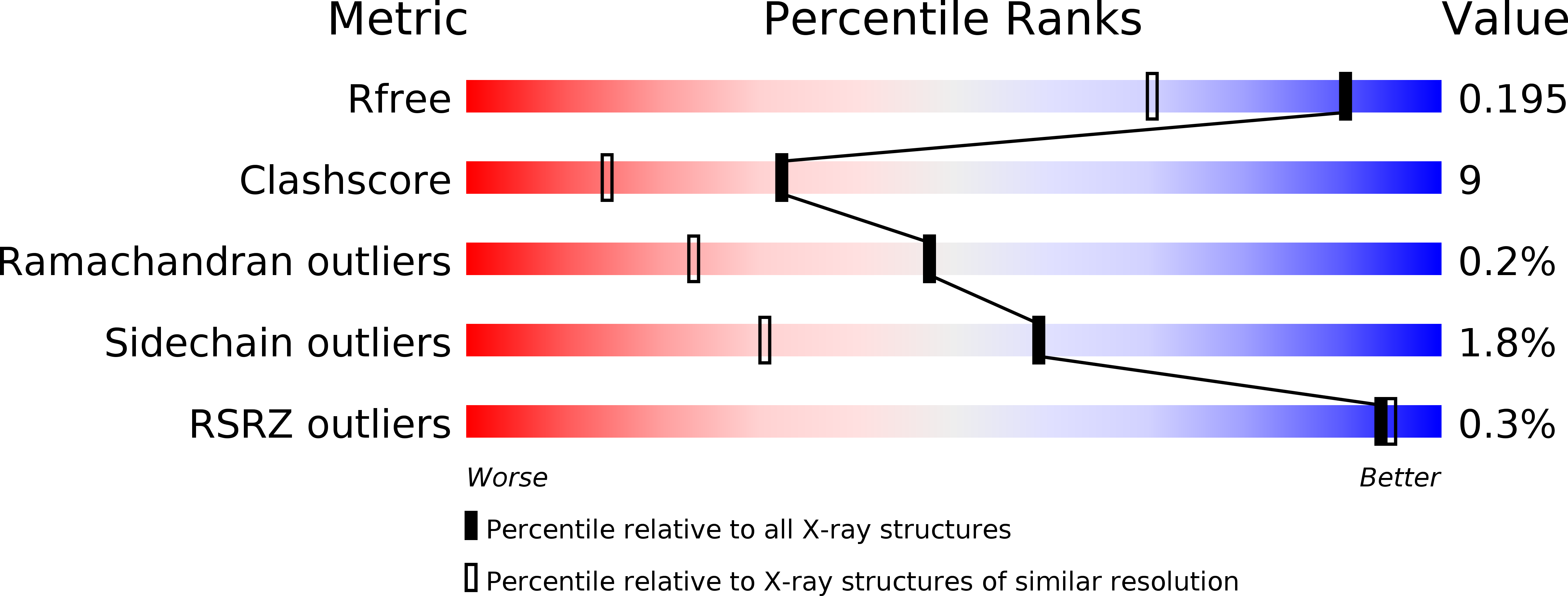
Resolution:
1.50 Å
R-Value Free:
0.19
R-Value Work:
0.15
R-Value Observed:
0.16
Space Group:
I 4
Macromolecular Entities
Polymer Type:polypeptide(L)
Description:Flavocytochrome c heme subunit
Chain IDs:A
Chain Length:174
Number of Molecules:1
Biological Source:Thermochromatium tepidum
Polymer Type:polypeptide(L)
Description:Flavocytochrome c flavin subunit
Chain IDs:B
Chain Length:401
Number of Molecules:1
Biological Source:Thermochromatium tepidum
Modified Residue
Compound ID Chain ID Parent Comp ID Details 2D Image
CSS B CYS S-MERCAPTOCYSTEINE
Primary Citation
Structure Analysis and Comparative Characterization of the Cytochrome c' and Flavocytochrome c from Thermophilic Purple Photosynthetic Bacterium Thermochromatium tepidum
Biochemistry 51 6556 6567 (2012)
PMID: 22827326 DOI: 10.1021/bi3005522

Abstact

The thermodynamic and spectroscopic properties of two soluble electron transport proteins, cytochrome (Cyt) c' and flavocytochrome c, isolated from thermophilic purple sulfur bacterium Thermochromatium (Tch.) tepidum were examined and compared with those of the corresponding proteins from a closely related mesophilic bacterium Allochromatium (Alc.) vinosum. These proteins share sequence identities of 82% for the cytochromes c' and 86% for the flavocytochromes c. Crystal structures of the two proteins have been determined at high resolutions. Differential scanning calorimetry and denaturing experiments show that both proteins from Tch. tepidum are thermally and structurally much more stable than their mesophilic counterparts. The denaturation temperature of Tch. tepidum Cyt c' was 22 °C higher than that of Alc. vinosum Cyt c', and the midpoints of denaturation using guanidine hydrochloride were 2.0 and 1.2 M for the Tch. tepidum and Alc. vinosum flavocytochromes c, respectively. The enhanced stabilities can be interpreted on the basis of the structural and sequence information obtained in this study: increased number of hydrogen bonds formed between main chain nitrogen and oxygen atoms, more compact structures and reduced number of glycine residues. Many residues with large side chains in Alc. vinosum Cyt c' are substituted by alanines in Tch. tepidum Cyt c'. Both proteins from Tch. tepidum exhibit high structural similarities to their counterparts from Alc. vinosum, and the different residues between the corresponding proteins are mainly located on the surface and exposed to the solvent. Water molecules are found in the heme vicinity of Tch. tepidum Cyt c' and form hydrogen bonds with the heme ligand and C-terminal charged residues. Similar bound waters are also found in the vicinity of one heme group in the diheme subunit of Tch. tepidum flavocytochrome c. Electron density map of the Tch. tepidum flavocytochrome c clearly revealed the presence of disulfur atoms positioned between two cysteine residues at the active site near the FAD prosthetic group. The result strongly suggests that flavocytochrome c is involved in the sulfide oxidation in vivo. Detailed discussion is given on the relationships between the crystal structures and the spectroscopic properties observed for these proteins.

Legend

Protein

Chemical

Disease

Primary Citation of related structures