3VR3 image
Deposition Date 2012-04-03
Release Date 2013-01-16
Last Version Date 2024-11-20
Entry Detail
PDB ID:
3VR3
Keywords:
Title:
Crystal structure of AMP-PNP bound A3B3 complex from Enterococcus hirae V-ATPase [bA3B3]
Biological Source:
Source Organism:
Host Organism:
Method Details:
Experimental Method:
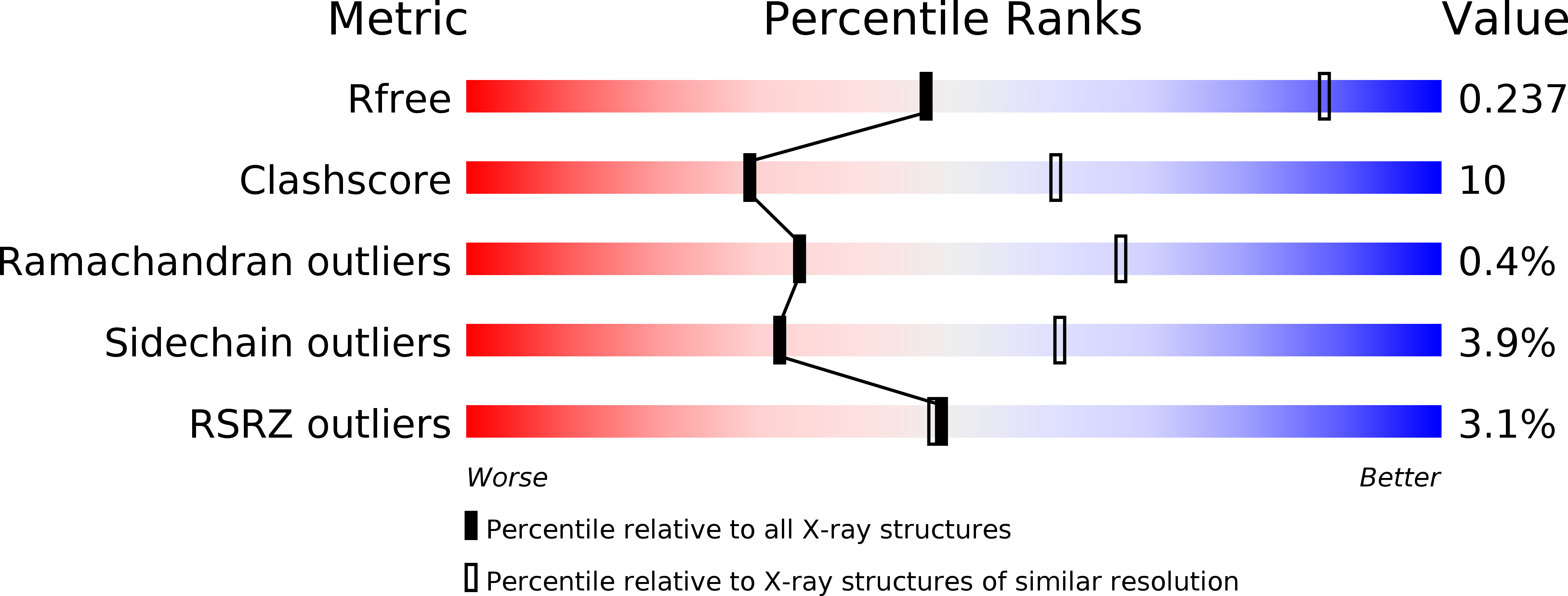
Resolution:
3.40 Å
R-Value Free:
0.23
R-Value Work:
0.19
R-Value Observed:
0.19
Space Group:
P 21 21 21
Macromolecular Entities
Polymer Type:polypeptide(L)
Molecule:V-type sodium ATPase catalytic subunit A
Gene (Uniprot):ntpA
Chain IDs:A, B, C
Chain Length:600
Number of Molecules:3
Biological Source:Enterococcus hirae
Polymer Type:polypeptide(L)
Molecule:V-type sodium ATPase subunit B
Gene (Uniprot):ntpB
Chain IDs:D, E, F
Chain Length:465
Number of Molecules:3
Biological Source:Enterococcus hirae
Modified Residue
Compound ID Chain ID Parent Comp ID Details 2D Image
MSE A MET SELENOMETHIONINE
Primary Citation
Rotation mechanism of Enterococcus hirae V(1)-ATPase based on asymmetric crystal structures
Nature 493 703 707 (2013)
PMID: 23334411 DOI: 10.1038/nature11778

Abstact

In various cellular membrane systems, vacuolar ATPases (V-ATPases) function as proton pumps, which are involved in many processes such as bone resorption and cancer metastasis, and these membrane proteins represent attractive drug targets for osteoporosis and cancer. The hydrophilic V(1) portion is known as a rotary motor, in which a central axis DF complex rotates inside a hexagonally arranged catalytic A(3)B(3) complex using ATP hydrolysis energy, but the molecular mechanism is not well defined owing to a lack of high-resolution structural information. We previously reported on the in vitro expression, purification and reconstitution of Enterococcus hirae V(1)-ATPase from the A(3)B(3) and DF complexes. Here we report the asymmetric structures of the nucleotide-free (2.8 Å) and nucleotide-bound (3.4 Å) A(3)B(3) complex that demonstrate conformational changes induced by nucleotide binding, suggesting a binding order in the right-handed rotational orientation in a cooperative manner. The crystal structures of the nucleotide-free (2.2 Å) and nucleotide-bound (2.7 Å) V(1)-ATPase are also reported. The more tightly packed nucleotide-binding site seems to be induced by DF binding, and ATP hydrolysis seems to be stimulated by the approach of a conserved arginine residue. To our knowledge, these asymmetric structures represent the first high-resolution view of the rotational mechanism of V(1)-ATPase.

Legend

Protein

Chemical

Disease

Primary Citation of related structures