Deposition Date
2012-04-02
Release Date
2013-01-02
Last Version Date
2023-11-08
Entry Detail
PDB ID:
3VQX
Keywords:
Title:
Crystal structure of the catalytic domain of pyrrolysyl-tRNA synthetase in triclinic crystal form
Biological Source:
Source Organism(s):
Methanosarcina mazei (Taxon ID: 2209)
Expression System(s):
Method Details:
Experimental Method:
Resolution:
2.30 Å
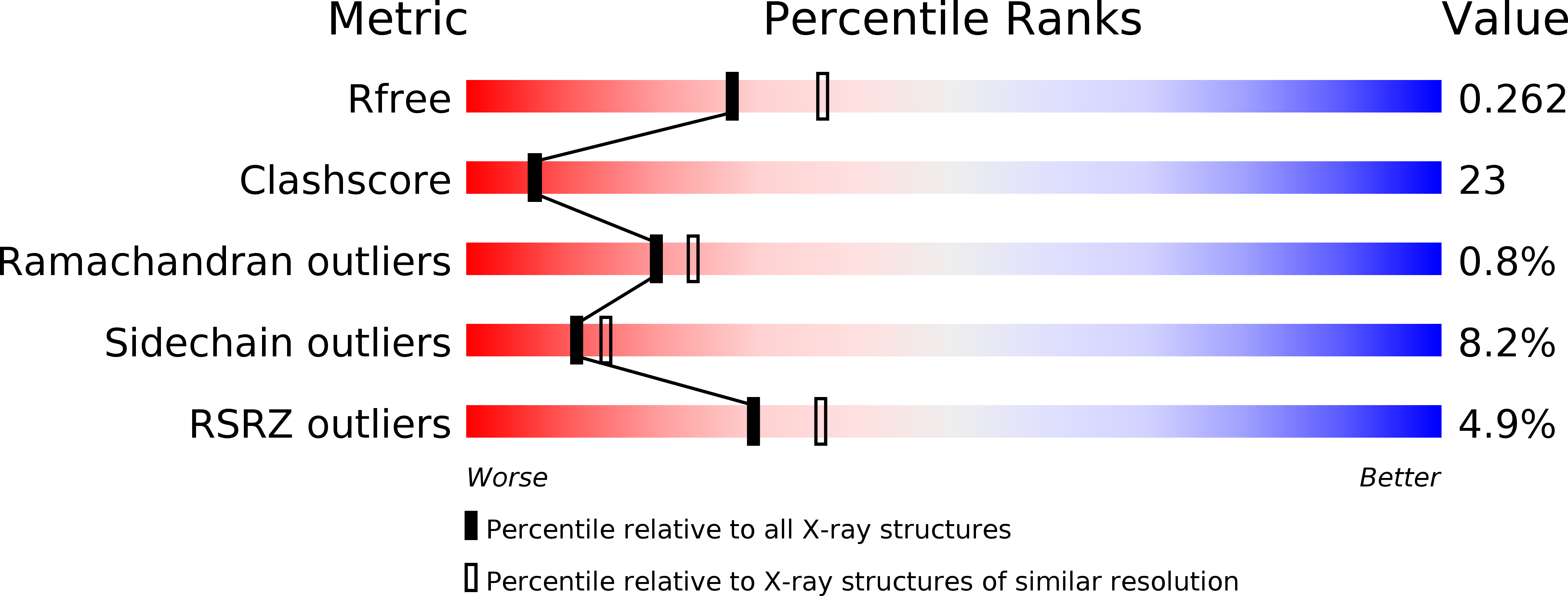
R-Value Free:
0.27
R-Value Work:
0.21
R-Value Observed:
0.21
Space Group:
P 1