3VQR image
Deposition Date 2012-03-29
Release Date 2012-04-25
Last Version Date 2023-11-08
Entry Detail
PDB ID:
3VQR
Keywords:
Title:
Structure of a dye-linked L-proline dehydrogenase mutant from the aerobic hyperthermophilic archaeon, Aeropyrum pernix
Biological Source:
Source Organism:
Host Organism:
Method Details:
Experimental Method:
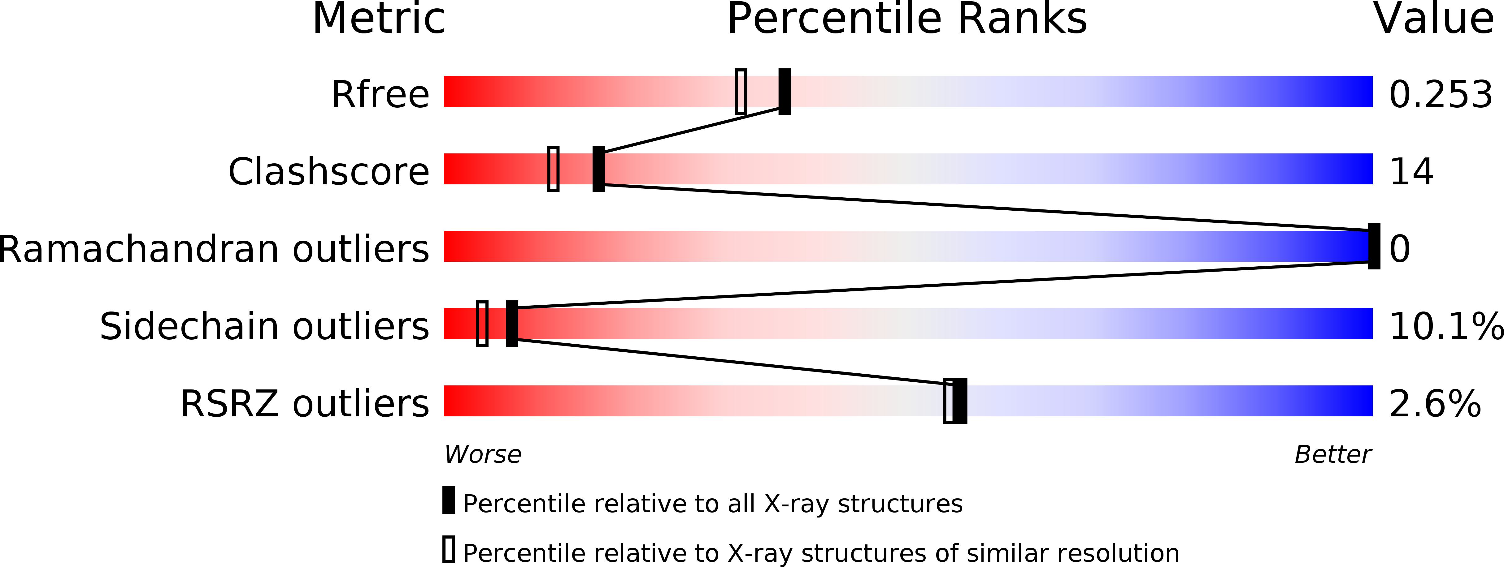
Resolution:
2.01 Å
R-Value Free:
0.25
R-Value Work:
0.21
R-Value Observed:
0.21
Space Group:
P 1
Macromolecular Entities
Polymer Type:polypeptide(L)
Molecule:Putative oxidoreductase
Gene (Uniprot):APE_1267.1
Mutations:delta L428
Chain IDs:A, B
Chain Length:447
Number of Molecules:2
Biological Source:Aeropyrum pernix
Primary Citation
Crystal Structure of Novel Dye-linked L-Proline Dehydrogenase from Hyperthermophilic Archaeon Aeropyrum pernix
J.Biol.Chem. 287 20070 20080 (2012)
PMID: 22511758 DOI: 10.1074/jbc.M111.319038

Abstact

Two types of dye-linked L-proline dehydrogenase (PDH1, α4β4-type hetero-octamer, and PDH2, αβγδ-type heterotetramer) have been identified so far in hyperthermophilic archaea. Here, we report the crystal structure of a third type of L-proline dehydrogenase, found in the aerobic hyperthermophilic archaeon Aeropyrum pernix, whose structure (homodimer) is much simpler than those of previously studied L-proline dehydrogenases. The structure was determined at a resolution of 1.92 Å. The asymmetric unit contained one subunit, and a crystallographic 2-fold axis generated the functional dimer. The overall fold of the subunit showed similarity to that of the PDH1 β-subunit, which is responsible for catalyzing L-proline dehydrogenation. However, the situation at the subunit-subunit interface of the A. pernix enzyme was totally different from that in PDH1. The presence of additional surface elements in the A. pernix enzyme contributes to a unique dimer association. Moreover, the C-terminal Leu(428), which is provided by a tail extending from the FAD-binding domain, shielded the active site, and an L-proline molecule was entrapped within the active site cavity. The K(m) value of a Leu(428) deletion mutant for L-proline was about 800 times larger than the K(m) value of the wild-type enzyme, although the k(cat) values did not differ much between the two enzymes. This suggests the C-terminal Leu(428) is not directly involved in catalysis, but it is essential for maintaining a high affinity for the substrate. This is the first description of an LPDH structure with L-proline bound, and it provides new insight into the substrate binding of LPDH.

Legend

Protein

Chemical

Disease

Primary Citation of related structures