3VQI image
Deposition Date 2012-03-24
Release Date 2012-08-01
Last Version Date 2023-11-08
Entry Detail
PDB ID:
3VQI
Title:
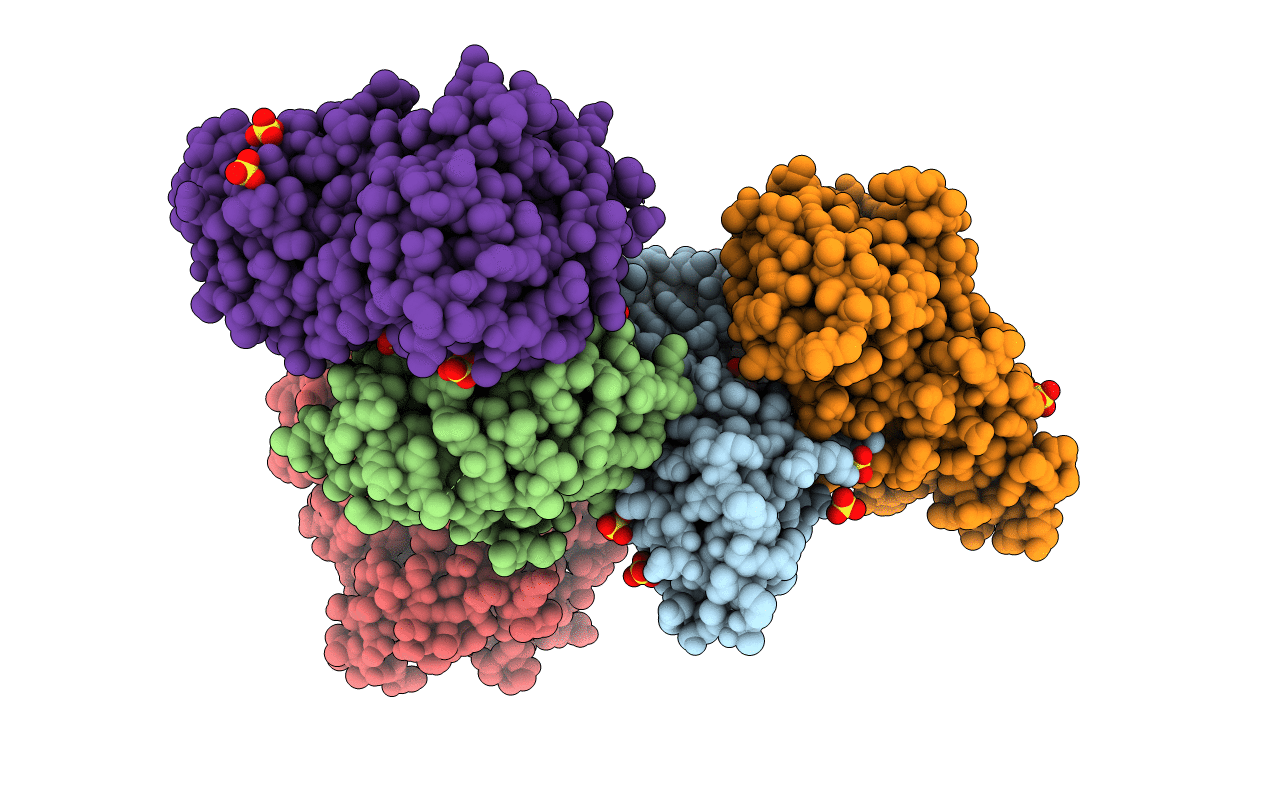
Crystal structure of Kluyveromyces marxianus Atg5
Biological Source:
Source Organism:
Host Organism:
Method Details:
Experimental Method:
Resolution:
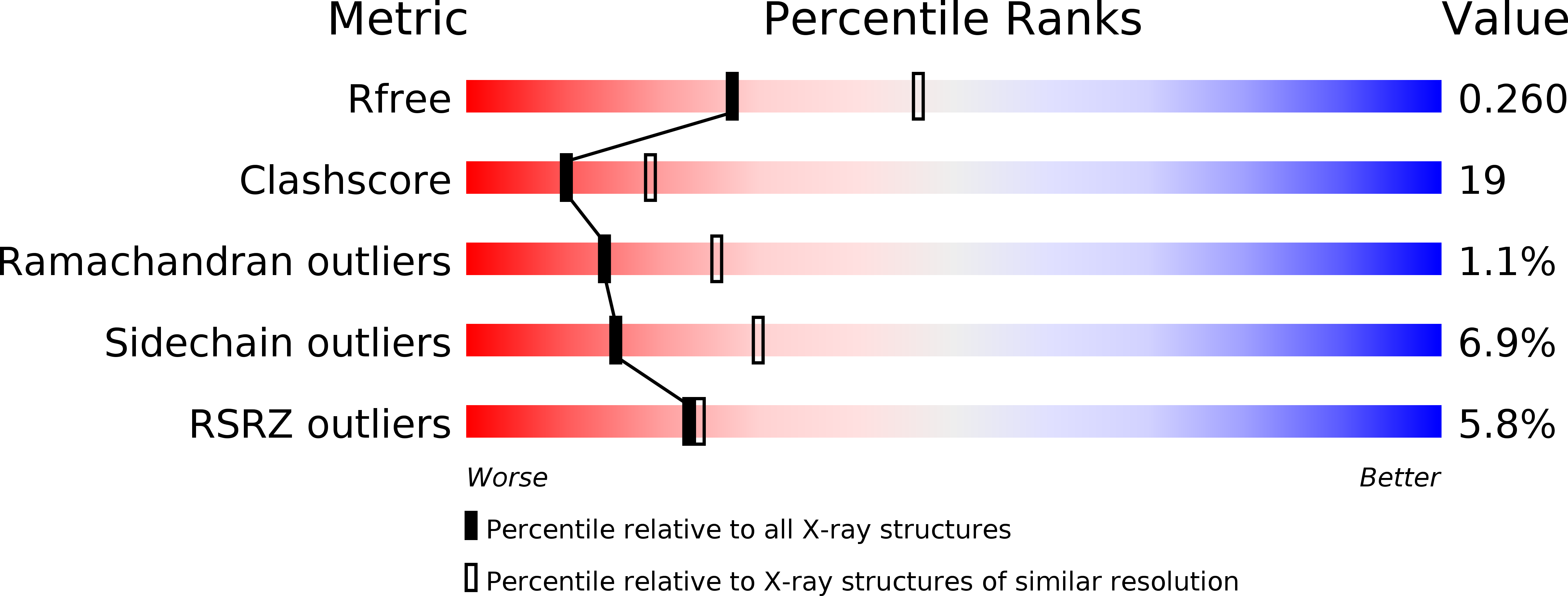
2.50 Å
R-Value Free:
0.26
R-Value Work:
0.23
R-Value Observed:
0.23
Space Group:
C 1 2 1
Macromolecular Entities
Polymer Type:polypeptide(L)
Molecule:Atg5
Chain IDs:A, B, C, D, E
Chain Length:274
Number of Molecules:5
Biological Source:Kluyveromyces marxianus
Primary Citation
Structural insights into atg10-mediated formation of the autophagy-essential atg12-atg5 conjugate
Structure 20 1244 1254 (2012)
PMID: 22682742 DOI: 10.1016/j.str.2012.04.018

Abstact

The Atg12-Atg5 conjugate, which is formed by an ubiquitin-like conjugation system, is essential to autophagosome formation, a central event in autophagy. Despite its importance, the molecular mechanism of the Atg12-Atg5 conjugate formation has not been elucidated. Here, we report the solution and crystal structures of Atg10 and Atg5 homologs from Kluyveromyces marxianus (Km), a thermotolerant yeast. KmAtg10 comprises an E2-core fold with characteristic accessories, including two β strands, whereas KmAtg5 has two ubiquitin-like domains and a helical domain. The nuclear magnetic resonance experiments, mutational analyses, and crosslinking experiments showed that KmAtg10 directly recognizes KmAtg5, especially its C-terminal ubiquitin-like domain, by its characteristic two β strands. Kinetic analysis suggests that Tyr56 and Asn114 of KmAtg10 may place the side chain of KmAtg5 Lys145 into the optimal orientation for its conjugation reaction with Atg12. These structural features enable Atg10 to mediate the formation of the Atg12-Atg5 conjugate without a specific E3 enzyme.

Legend

Protein

Chemical

Disease

Primary Citation of related structures