3VPK image
Deposition Date 2012-03-05
Release Date 2012-05-23
Last Version Date 2024-11-20
Entry Detail
PDB ID:
3VPK
Title:
Crystal Structure of 6-Guanidinohexanoyl Trypsin
Biological Source:
Source Organism:
Bos taurus (Taxon ID: 9913)
Method Details:
Experimental Method:
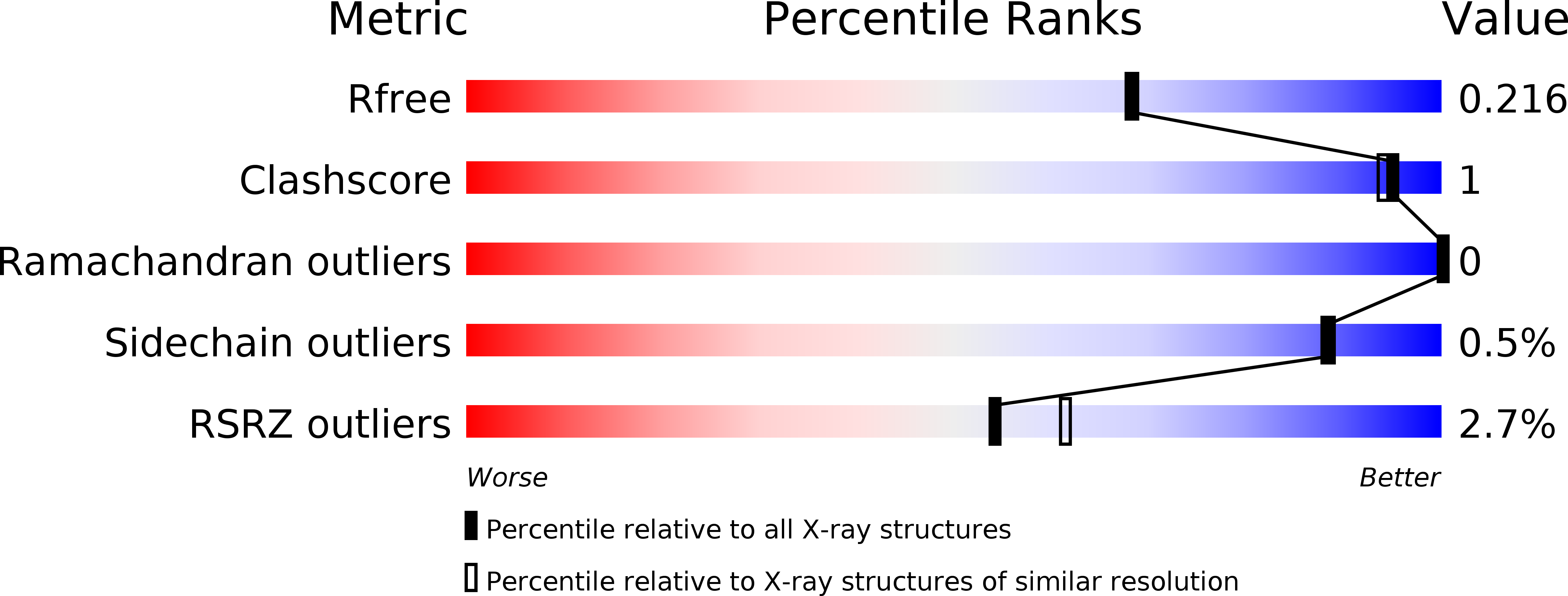
Resolution:
1.94 Å
R-Value Free:
0.21
R-Value Work:
0.18
R-Value Observed:
0.18
Space Group:
P 21 21 21
Macromolecular Entities
Structures with similar UniProt ID
Protein Blast
Polymer Type:polypeptide(L)
Molecule:Cationic trypsin
Gene (Uniprot):PRSS1
Chain IDs:A
Chain Length:223
Number of Molecules:1
Biological Source:Bos taurus
Primary Citation
Crystal structure of 6-guanidinohexanoyl trypsin near the optimum pH reveals the acyl-enzyme intermediate to be deacylated
Proteins ? ? ? (2012)
PMID: 23161653 DOI: 10.1002/prot.24206

Abstact

The force driving the conversion from the acyl intermediate to the tetrahedral intermediate in the deacylation reaction of serine proteases remains unclear. The crystal structure of 6-guanidinohexanoyl trypsin was determined at pH 7.0, near the optimum reaction pH, at 1.94 Å resolution. In this structure, three water molecules are observed around the catalytic site. One acts as a nucleophile to attack the acyl carbonyl carbon while the other two waters fix the position of the catalytic water through a hydrogen bond. When the acyl carbonyl oxygen oscillates thermally, the water assumes an appropriate angle to catalyze the deacylation.

Legend

Protein

Chemical

Disease

Primary Citation of related structures