Deposition Date
2012-02-22
Release Date
2012-10-03
Last Version Date
2023-11-08
Entry Detail
PDB ID:
3VOW
Keywords:
Title:
Crystal Structure of the Human APOBEC3C having HIV-1 Vif-binding Interface
Biological Source:
Source Organism(s):
Homo sapiens (Taxon ID: 9606)
Expression System(s):
Method Details:
Experimental Method:
Resolution:
2.15 Å
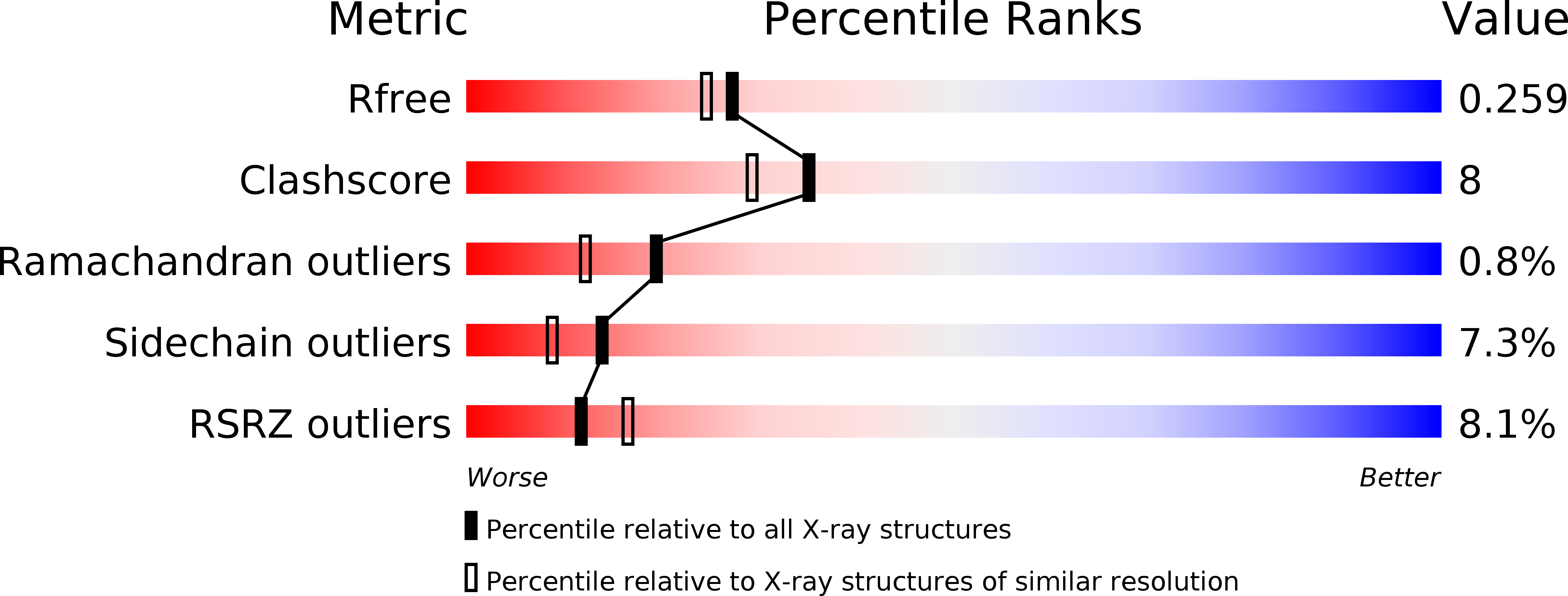
R-Value Free:
0.26
R-Value Work:
0.21
R-Value Observed:
0.21
Space Group:
P 61