
Deposition Date
2011-12-09
Release Date
2012-03-14
Last Version Date
2023-11-08
Entry Detail
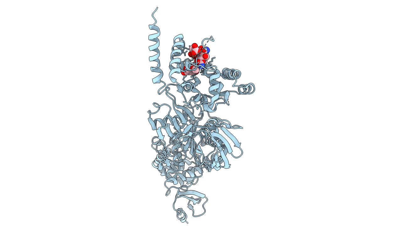
PDB ID:
3VMA
Keywords:
Title:
Crystal Structure of the Full-Length Transglycosylase PBP1b from Escherichia coli
Biological Source:
Source Organism(s):
Escherichia coli (Taxon ID: 83333)
Expression System(s):
Method Details:
Experimental Method:
Resolution:
2.16 Å
R-Value Free:
0.25
R-Value Work:
0.21
R-Value Observed:
0.21
Space Group:
P 21 21 2
