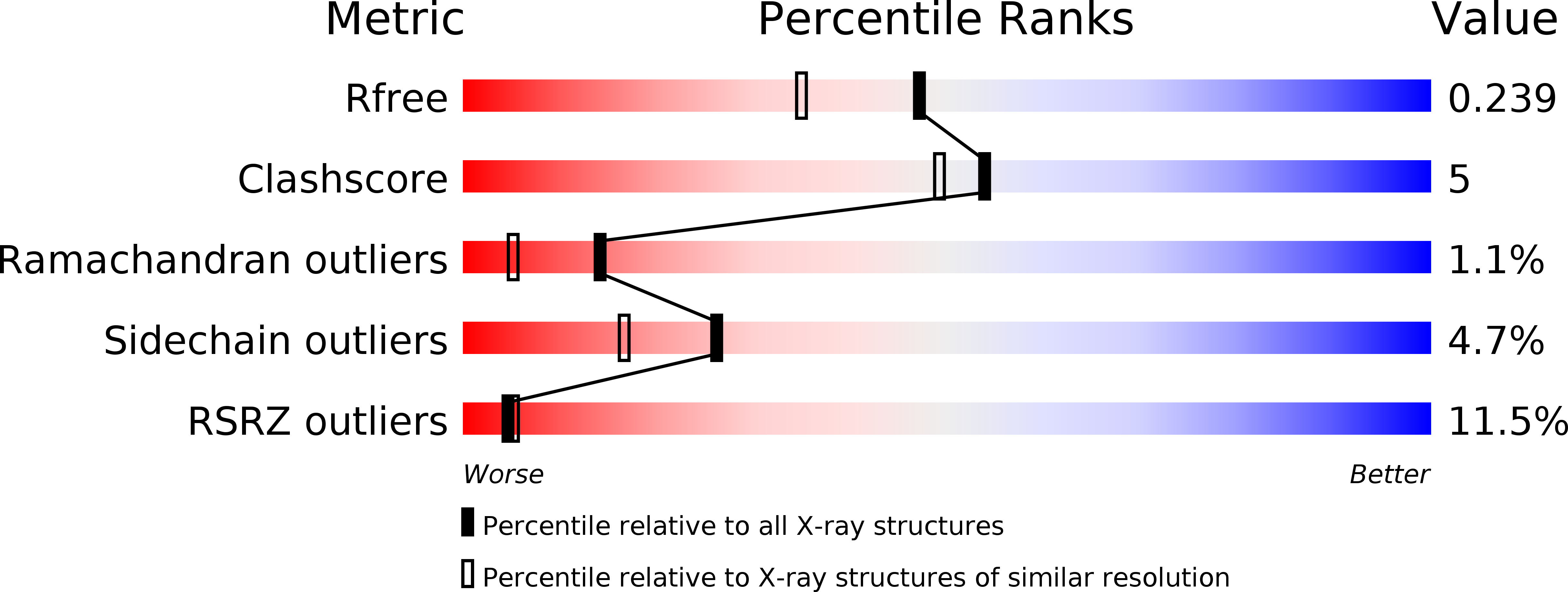
3VK6 image
Deposition Date 2011-11-09
Release Date 2012-01-25
Last Version Date 2024-03-20
Entry Detail
PDB ID:
3VK6
Keywords:
Title:
Crystal structure of a phosphotyrosine binding domain
Biological Source:
Source Organism:
Mus musculus (Taxon ID: 10090)
Host Organism:
Method Details:
Experimental Method:
Resolution:
1.90 Å
R-Value Free:
0.24
R-Value Work:
0.21
R-Value Observed:
0.22
Space Group:
P 62 2 2
Macromolecular Entities
Polymer Type:polypeptide(L)
Molecule:E3 ubiquitin-protein ligase Hakai
Gene (Uniprot):Cbll1
Chain IDs:A
Chain Length:101
Number of Molecules:1
Biological Source:Mus musculus
Ligand Molecules
Primary Citation
Structure of a novel phosphotyrosine-binding domain in Hakai that targets E-cadherin
Embo J. 31 1308 1319 (2012)
PMID: 22252131 DOI: 10.1038/emboj.2011.496

Abstact

Phosphotyrosine-binding domains, typified by the SH2 (Src homology 2) and PTB domains, are critical upstream components of signal transduction pathways. The E3 ubiquitin ligase Hakai targets tyrosine-phosphorylated E-cadherin via an uncharacterized domain. In this study, the crystal structure of Hakai (amino acids 106-206) revealed that it forms an atypical, zinc-coordinated homodimer by utilizing residues from the phosphotyrosine-binding domain of two Hakai monomers. Hakai dimerization allows the formation of a phosphotyrosine-binding pocket that recognizes specific phosphorylated tyrosines and flanking acidic amino acids of Src substrates, such as E-cadherin, cortactin and DOK1. NMR and mutational analysis identified the Hakai residues required for target binding within the binding pocket, now named the HYB domain. ZNF645 also possesses a HYB domain but demonstrates different target specificities. The HYB domain is structurally different from other phosphotyrosine-binding domains and is a potential drug target due to its novel structural features.

Legend

Protein

Chemical

Disease

Primary Citation of related structures