
Deposition Date
2011-09-28
Release Date
2012-06-06
Last Version Date
2024-10-30
Entry Detail
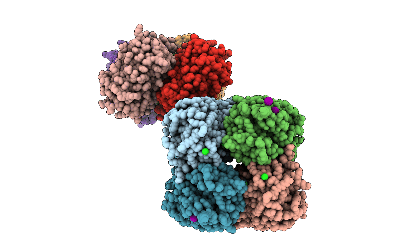
PDB ID:
3VIC
Keywords:
Title:
Green-fluorescent variant of the non-fluorescent chromoprotein Rtms5
Biological Source:
Source Organism(s):
Montipora efflorescens (Taxon ID: 105610)
Expression System(s):
Method Details:
Experimental Method:
Resolution:
2.20 Å
R-Value Free:
0.20
R-Value Work:
0.15
R-Value Observed:
0.16
Space Group:
C 2 2 21
