3VHJ image
Deposition Date 2011-08-25
Release Date 2012-05-23
Last Version Date 2024-10-30
Entry Detail
PDB ID:
3VHJ
Title:
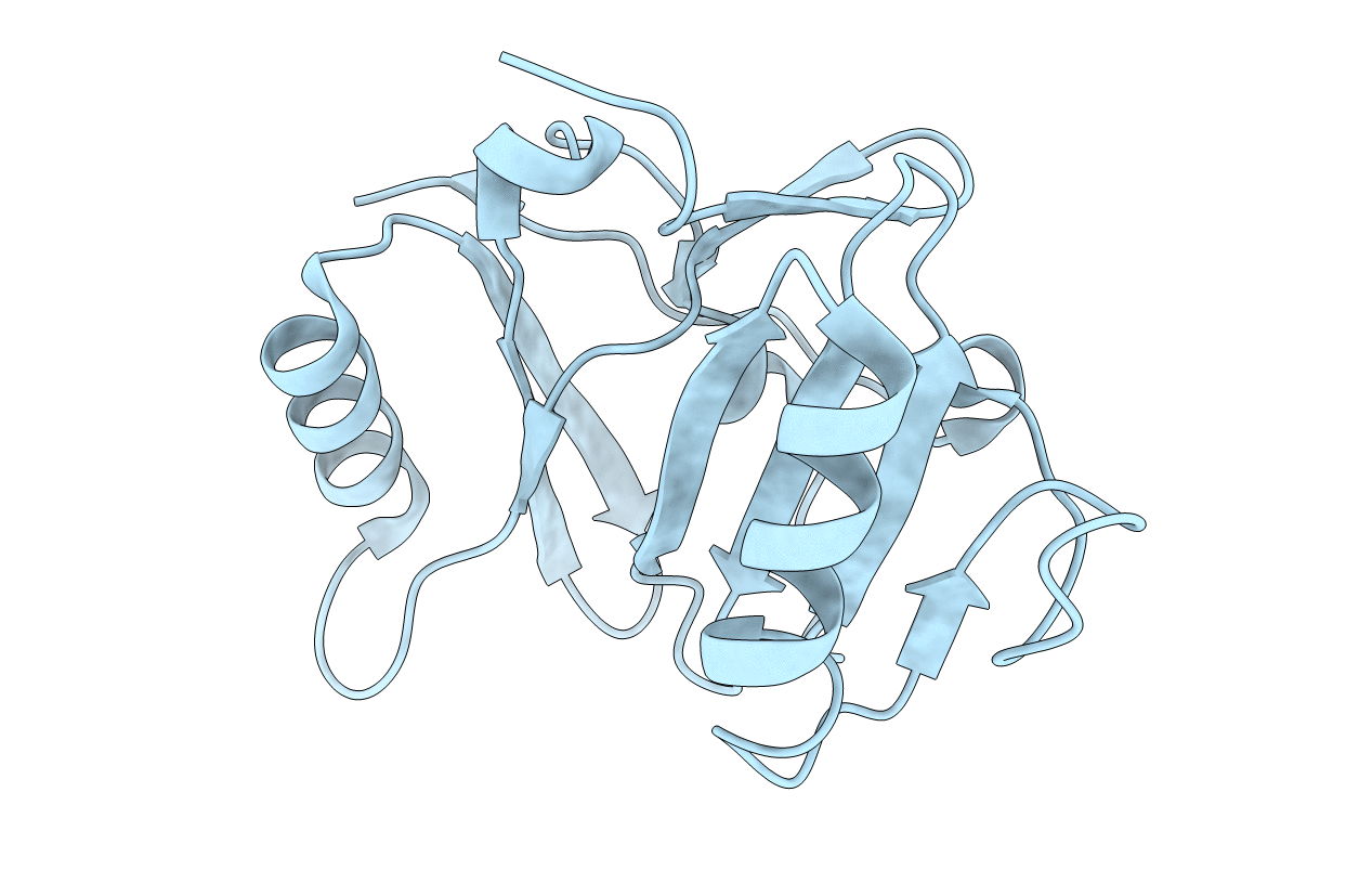
Crystal structure of the cytoplasmic domain of BfpC
Biological Source:
Source Organism:
Escherichia coli (Taxon ID: 562)
Host Organism:
Method Details:
Experimental Method:
Resolution:
1.90 Å
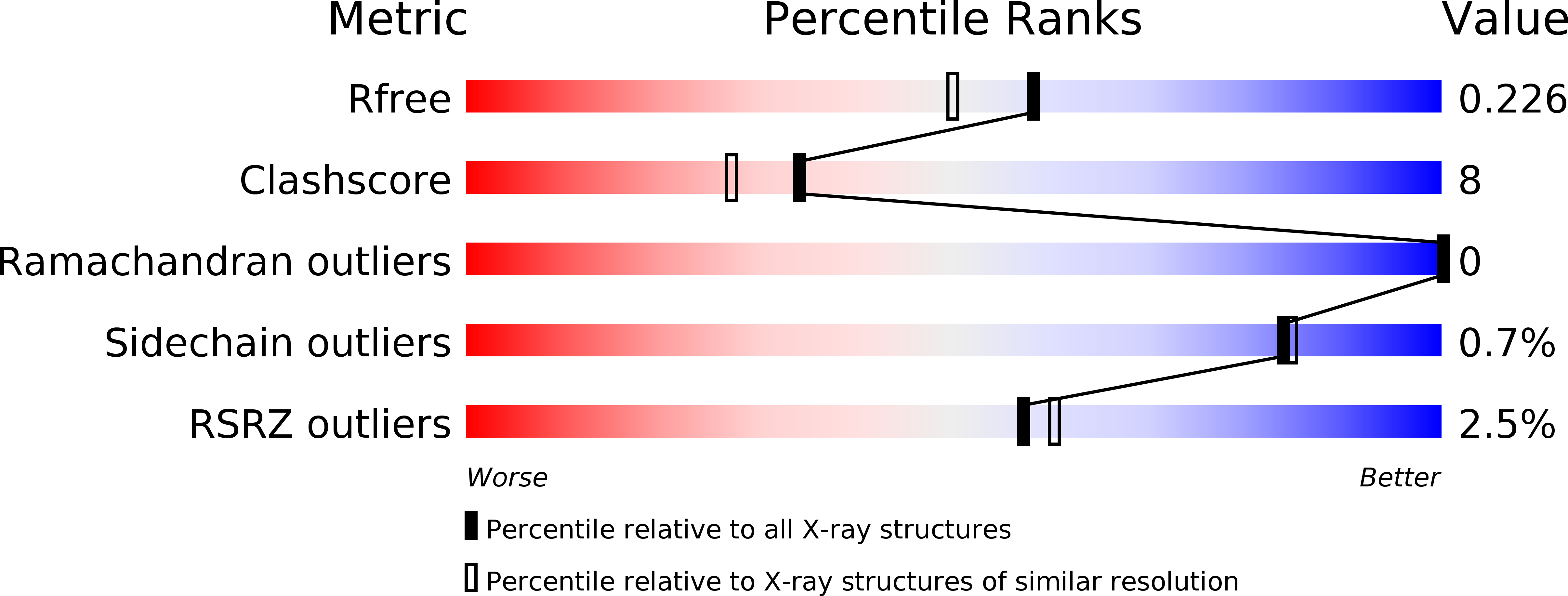
R-Value Free:
0.22
R-Value Work:
0.21
R-Value Observed:
0.21
Space Group:
P 21 21 21
Macromolecular Entities
Polymer Type:polypeptide(L)
Molecule:BfpC
Mutations:I2V
Chain IDs:A
Chain Length:172
Number of Molecules:1
Biological Source:Escherichia coli
Modified Residue
Compound ID Chain ID Parent Comp ID Details 2D Image
MSE A MET SELENOMETHIONINE
Primary Citation
Structure of an Essential Type IV Pilus Biogenesis Protein Provides Insights into Pilus and Type II Secretion Systems
J.Mol.Biol. 419 110 124 (2012)
PMID: 22387466 DOI: 10.1016/j.jmb.2012.02.041

Abstact

Type IV pili (T4Ps) are long cell surface filaments, essential for microcolony formation, tissue adherence, motility, transformation, and virulence by human pathogens. The enteropathogenic Escherichia coli bundle-forming pilus is a prototypic T4P assembled and powered by BfpD, a conserved GspE secretion superfamily ATPase held by inner-membrane proteins BfpC and BfpE, a GspF-family membrane protein. Although the T4P assembly machinery shares similarity with type II secretion (T2S) systems, the structural biochemistry of the T4P machine has been obscure. Here, we report the crystal structure of the two-domain BfpC cytoplasmic region (N-BfpC), responsible for binding to ATPase BfpD and membrane protein BfpE. The N-BfpC structure reveals a prominent central cleft between two α/β-domains. Despite negligible sequence similarity, N-BfpC resembles PilM, a cytoplasmic T4P biogenesis protein. Yet surprisingly, N-BfpC has far greater structural similarity to T2S component EpsL, with which it also shares virtually no sequence identity. The C-terminus of the cytoplasmic domain, which leads to the transmembrane segment not present in the crystal structure, exits N-BfpC at a positively charged surface that most likely interacts with the inner membrane, positioning its central cleft for interactions with other Bfp components. Point mutations in surface-exposed N-BfpC residues predicted to be critical for interactions among BfpC, BfpE, and BfpD disrupt pilus biogenesis without precluding interactions with BfpE and BfpD and without affecting BfpD ATPase activity. These results illuminate the relationships between T4P biogenesis and T2S systems, imply that subtle changes in component residue interactions can have profound effects on function and pathogenesis, and suggest that T4P systems may be disrupted by inhibitors that do not preclude component assembly.

Legend

Protein

Chemical

Disease

Primary Citation of related structures