3VH2 image
Deposition Date 2011-08-23
Release Date 2011-09-21
Last Version Date 2023-11-08
Entry Detail
PDB ID:
3VH2
Title:
Crystal structure of Saccharomyces cerevisiae Atg7 (1-613)
Biological Source:
Source Organism:
Host Organism:
Method Details:
Experimental Method:
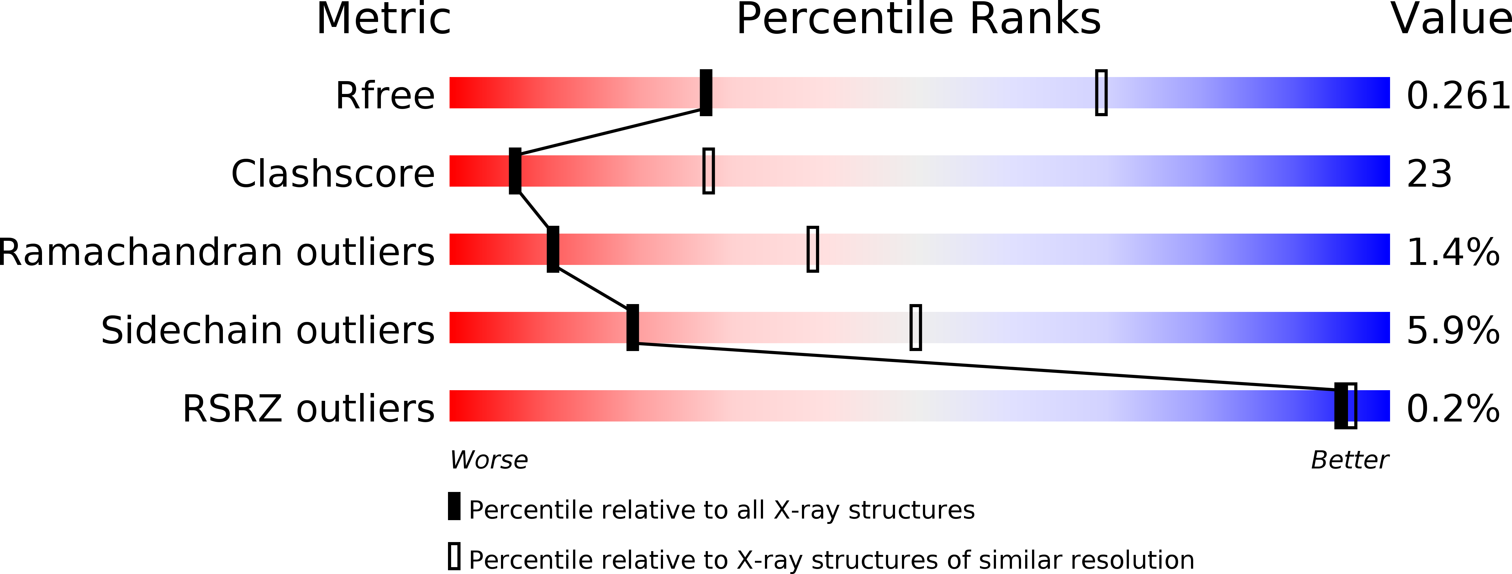
Resolution:
3.30 Å
R-Value Free:
0.26
R-Value Work:
0.21
R-Value Observed:
0.21
Space Group:
P 31 2 1
Macromolecular Entities
Polymer Type:polypeptide(L)
Molecule:Ubiquitin-like modifier-activating enzyme ATG7
Gene (Uniprot):ATG7
Chain IDs:A
Chain Length:616
Number of Molecules:1
Biological Source:Saccharomyces cerevisiae
Ligand Molecules
Primary Citation
Structural basis of Atg8 activation by a homodimeric E1, Atg7.
Mol.Cell 44 462 475 (2011)
PMID: 22055191 DOI: 10.1016/j.molcel.2011.08.035

Abstact

E1 enzymes activate ubiquitin-like proteins and transfer them to cognate E2 enzymes. Atg7, a noncanonical E1, activates two ubiquitin-like proteins, Atg8 and Atg12, and plays a crucial role in autophagy. Here, we report crystal structures of full-length Atg7 and its C-terminal domain bound to Atg8 and MgATP, as well as a solution structure of Atg8 bound to the extreme C-terminal domain (ECTD) of Atg7. The unique N-terminal domain (NTD) of Atg7 is responsible for Atg3 (E2) binding, whereas its C-terminal domain is comprised of a homodimeric adenylation domain (AD) and ECTD. The structural and biochemical data demonstrate that Atg8 is initially recognized by the C-terminal tail of ECTD and is then transferred to an AD, where the Atg8 C terminus is attacked by the catalytic cysteine to form a thioester bond. Atg8 is then transferred via a trans mechanism to the Atg3 bound to the NTD of the opposite protomer within a dimer.

Legend

Protein

Chemical

Disease

Primary Citation of related structures