3VGW image
Deposition Date 2011-08-21
Release Date 2011-12-28
Last Version Date 2024-11-20
Entry Detail
PDB ID:
3VGW
Title:
Crystal structure of monoAc-biotin-avidin complex
Biological Source:
Source Organism:
Gallus gallus (Taxon ID: 9031)
Method Details:
Experimental Method:
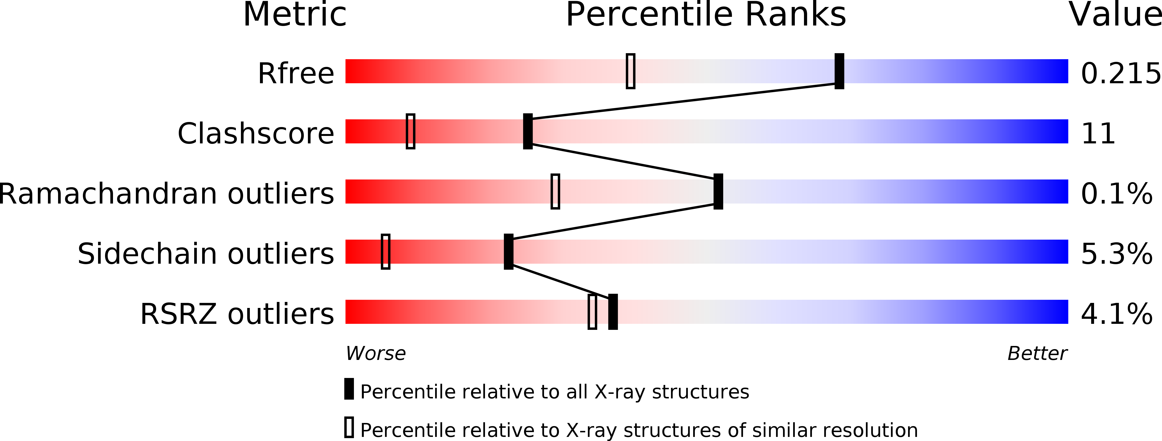
Resolution:
1.60 Å
R-Value Free:
0.21
R-Value Work:
0.18
R-Value Observed:
0.19
Space Group:
P 1 21 1
Macromolecular Entities
Polymer Type:polypeptide(L)
Molecule:Avidin
Gene (Uniprot):AVD
Chain IDs:A, B, C, D, E, F, G, H
Chain Length:123
Number of Molecules:8
Biological Source:Gallus gallus
Modified Residue
Compound ID Chain ID Parent Comp ID Details 2D Image
ASN A ASN GLYCOSYLATION SITE
Primary Citation
Rational development of caged-biotin protein-labeling agents and some applications in live cells
Chem.Biol. 18 1261 1272 (2011)
PMID: 22035795 DOI: 10.1016/j.chembiol.2011.09.007

Abstact

Biotin-(strept)avidin complex is widely used in biotechnology because of its extremely high binding constant, but there is no report describing spatiotemporally controlled formation of the complex in live cells. Here, based on X-ray crystal structure analysis and calorimetric data, we designed and synthesized photoreleasable biotins, which show greatly reduced affinity for (strept)avidin, but recover native affinity after UV irradiation. For application at the cell surface, we introduced an amine-reactive moiety into these "caged" biotin molecules. Specific fluorescence imaging of live cells that had been labeled with these agents and then UV-irradiated, was accomplished by addition of streptavidin conjugated with a fluorophore. We also demonstrated the applicability of these compounds for UV-irradiated-cell-specific drug delivery by using caged-biotin-labeled cells, a prodrug, and streptavidin conjugated with a prodrug-activating enzyme.

Legend

Protein

Chemical

Disease

Primary Citation of related structures