Deposition Date
2011-08-16
Release Date
2012-08-22
Last Version Date
2023-11-08
Entry Detail
PDB ID:
3VGN
Keywords:
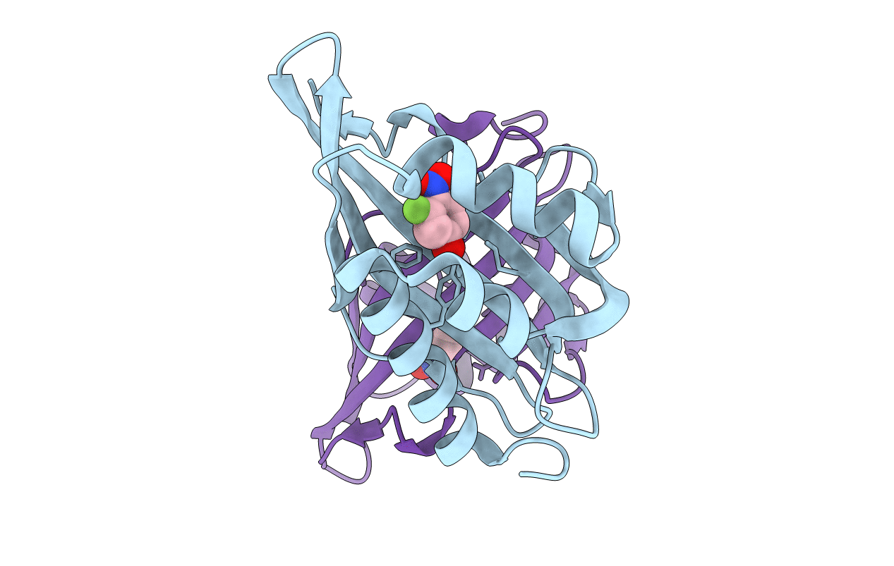
Title:
Crystal Structure of Ketosteroid Isomerase D40N from Pseudomonas putida (pKSI) with bound 3-fluoro-4-nitrophenol
Biological Source:
Source Organism(s):
Pseudomonas putida (Taxon ID: 303)
Expression System(s):
Method Details:
Experimental Method:
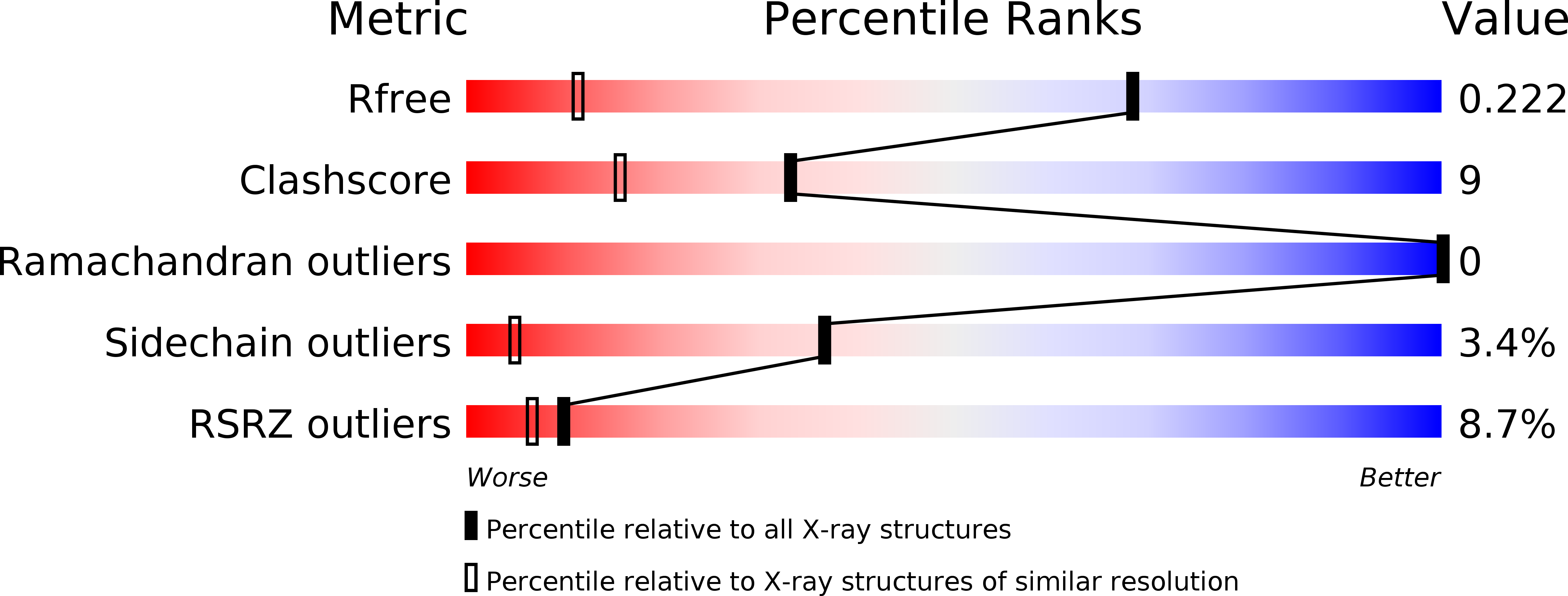
Resolution:
1.30 Å
R-Value Free:
0.21
R-Value Work:
0.16
R-Value Observed:
0.17
Space Group:
P 21 21 21