3VGF image
Deposition Date 2011-08-09
Release Date 2012-06-20
Last Version Date 2024-11-13
Entry Detail
PDB ID:
3VGF
Keywords:
Title:
Crystal structure of glycosyltrehalose trehalohydrolase (D252S) complexed with maltotriosyltrehalose
Biological Source:
Source Organism:
Host Organism:
Method Details:
Experimental Method:
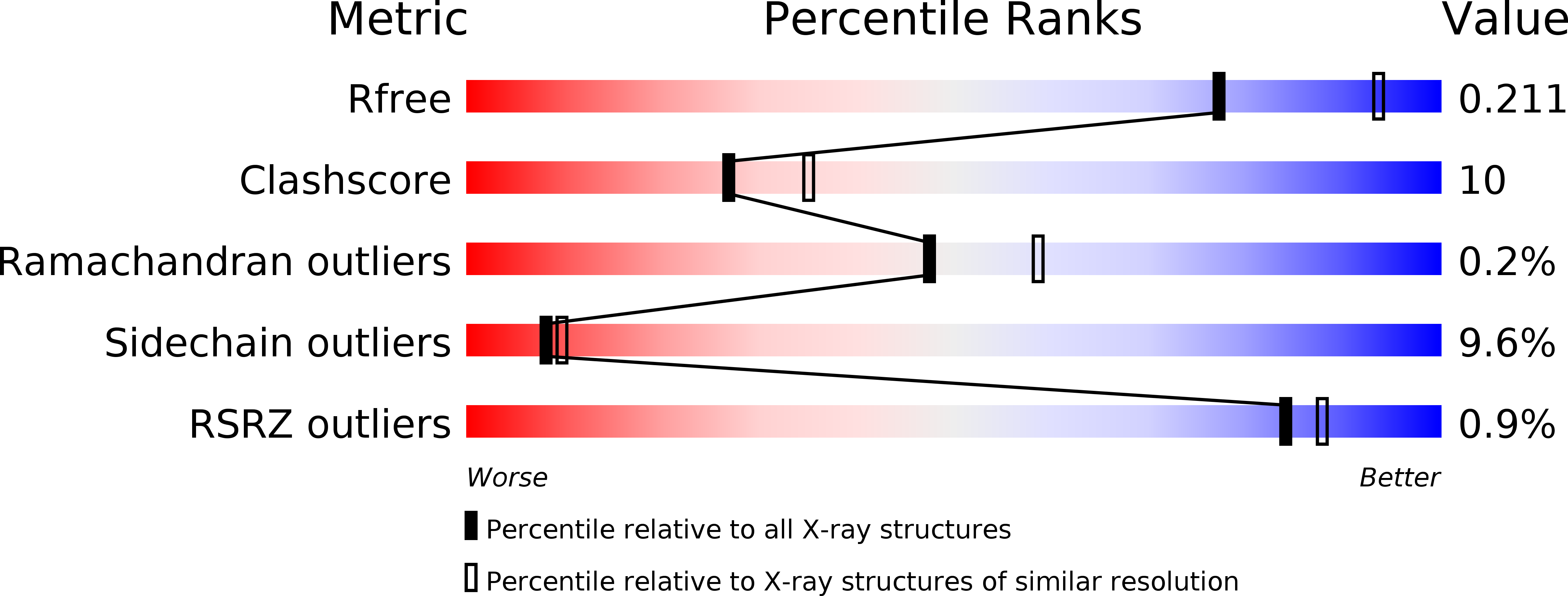
Resolution:
2.30 Å
R-Value Free:
0.21
R-Value Work:
0.17
R-Value Observed:
0.17
Space Group:
P 32 2 1
Macromolecular Entities
Polymer Type:polypeptide(L)
Molecule:Malto-oligosyltrehalose trehalohydrolase
Gene (Uniprot):treZ
Mutations:D252S
Chain IDs:A
Chain Length:558
Number of Molecules:1
Biological Source:Sulfolobus solfataricus
Peptide-like Molecules
PRD_900009
Primary Citation
Substrate recognition mechanism of a glycosyltrehalose trehalohydrolase from Sulfolobus solfataricus KM1.
Protein Sci. 21 539 552 (2012)
PMID: 22334583 DOI: 10.1002/pro.2039

Abstact

Glycosyltrehalose trehalohydrolase (GTHase) is an α-amylase that cleaves the α-1,4 bond adjacent to the α-1,1 bond of maltooligosyltrehalose to release trehalose. To investigate the catalytic and substrate recognition mechanisms of GTHase, two residues, Asp252 (nucleophile) and Glu283 (general acid/base), located at the catalytic site of GTHase were mutated (Asp252→Ser (D252S), Glu (D252E) and Glu283→Gln (E283Q)), and the activity and structure of the enzyme were investigated. The E283Q, D252E, and D252S mutants showed only 0.04, 0.03, and 0.6% of enzymatic activity against the wild-type, respectively. The crystal structure of the E283Q mutant GTHase in complex with the substrate, maltotriosyltrehalose (G3-Tre), was determined to 2.6-Å resolution. The structure with G3-Tre indicated that GTHase has at least five substrate binding subsites and that Glu283 is the catalytic acid, and Asp252 is the nucleophile that attacks the C1 carbon in the glycosidic linkage of G3-Tre. The complex structure also revealed a scheme for substrate recognition by GTHase. Substrate recognition involves two unique interactions: stacking of Tyr325 with the terminal glucose ring of the trehalose moiety and perpendicularly placement of Trp215 to the pyranose rings at the subsites -1 and +1 glucose.

Legend

Protein

Chemical

Disease

Primary Citation of related structures