Deposition Date
2011-08-04
Release Date
2012-02-01
Last Version Date
2024-10-30
Entry Detail
PDB ID:
3VGA
Keywords:
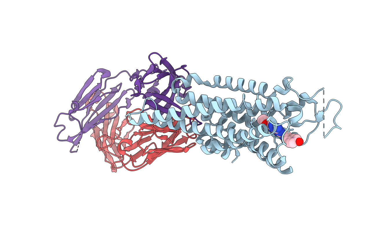
Title:
Crystal structure of human adenosine A2A receptor with an allosteric inverse-agonist antibody at 3.1 A resolution
Biological Source:
Source Organism(s):
Homo sapiens (Taxon ID: 9606)
Mus musculus (Taxon ID: 10090)
Mus musculus (Taxon ID: 10090)
Expression System(s):
Method Details:
Experimental Method:
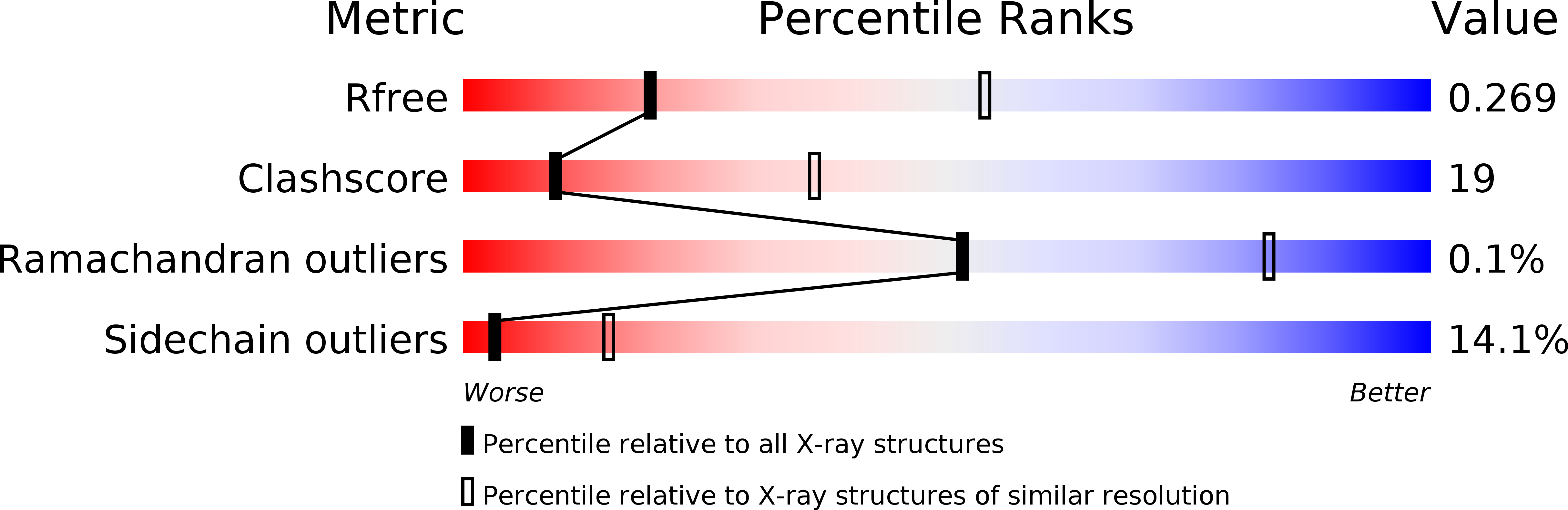
Resolution:
3.10 Å
R-Value Free:
0.26
R-Value Work:
0.19
R-Value Observed:
0.19
Space Group:
C 1 2 1