Deposition Date
2012-01-06
Release Date
2012-02-22
Last Version Date
2024-11-27
Entry Detail
PDB ID:
3VE1
Keywords:
Title:
The 2.9 angstrom crystal structure of Transferrin binding protein B (TbpB) from serogroup B M982 Neisseria meningitidis in complex with human transferrin
Biological Source:
Source Organism(s):
Neisseria meningitidis serogroup B (Taxon ID: 491)
Homo sapiens (Taxon ID: 9606)
Homo sapiens (Taxon ID: 9606)
Expression System(s):
Method Details:
Experimental Method:
Resolution:
2.96 Å
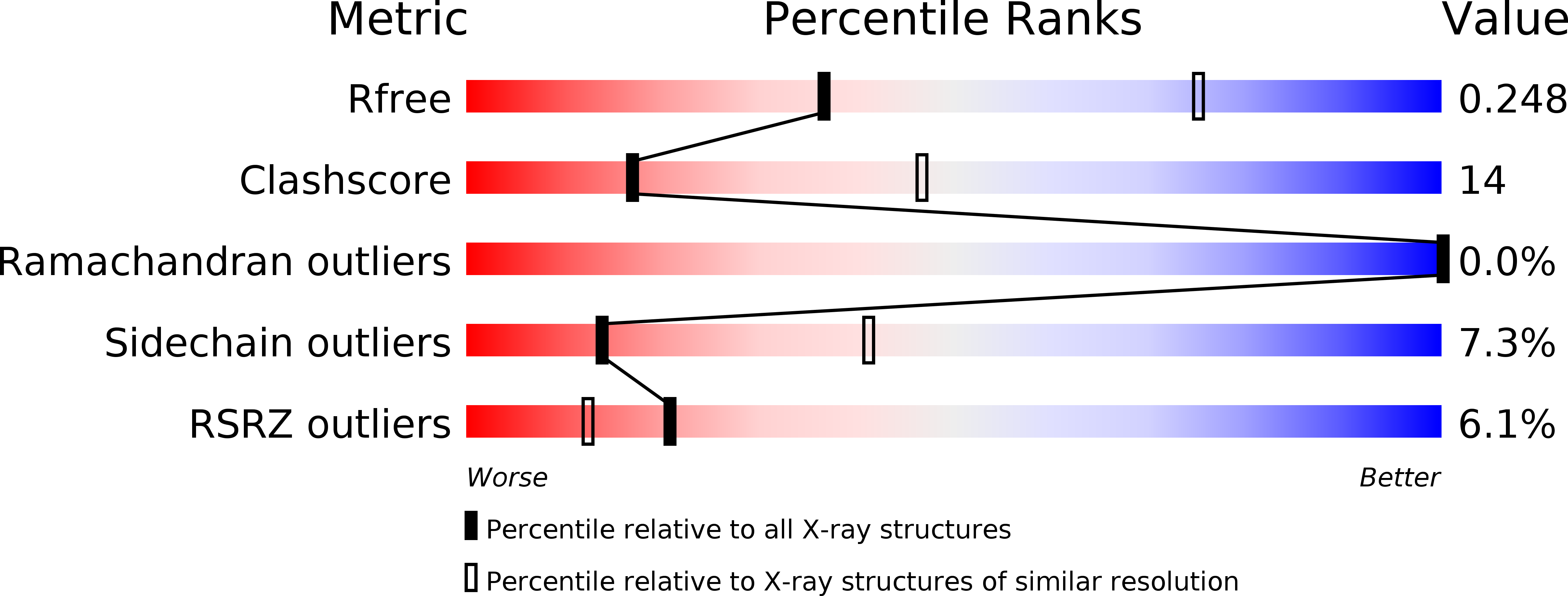
R-Value Free:
0.24
R-Value Work:
0.20
R-Value Observed:
0.20
Space Group:
P 21 21 21