Deposition Date
2012-01-06
Release Date
2012-11-28
Last Version Date
2024-02-28
Entry Detail
PDB ID:
3VDZ
Keywords:
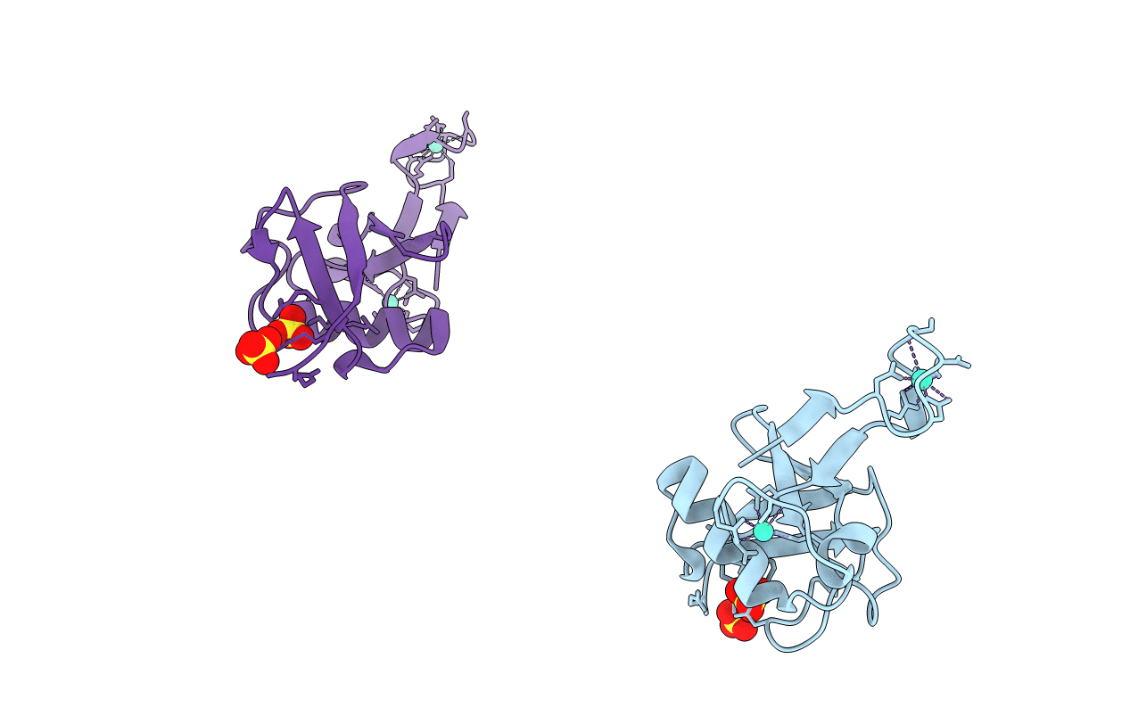
Title:
Tailoring Encodable Lanthanide-Binding Tags as MRI Contrast Agents: xq-dSE3-Ubiquitin at 2.4 Angstroms
Biological Source:
Source Organism(s):
synthetic construct, homo sapiens (Taxon ID: 32630, 9606)
Expression System(s):
Method Details:
Experimental Method:
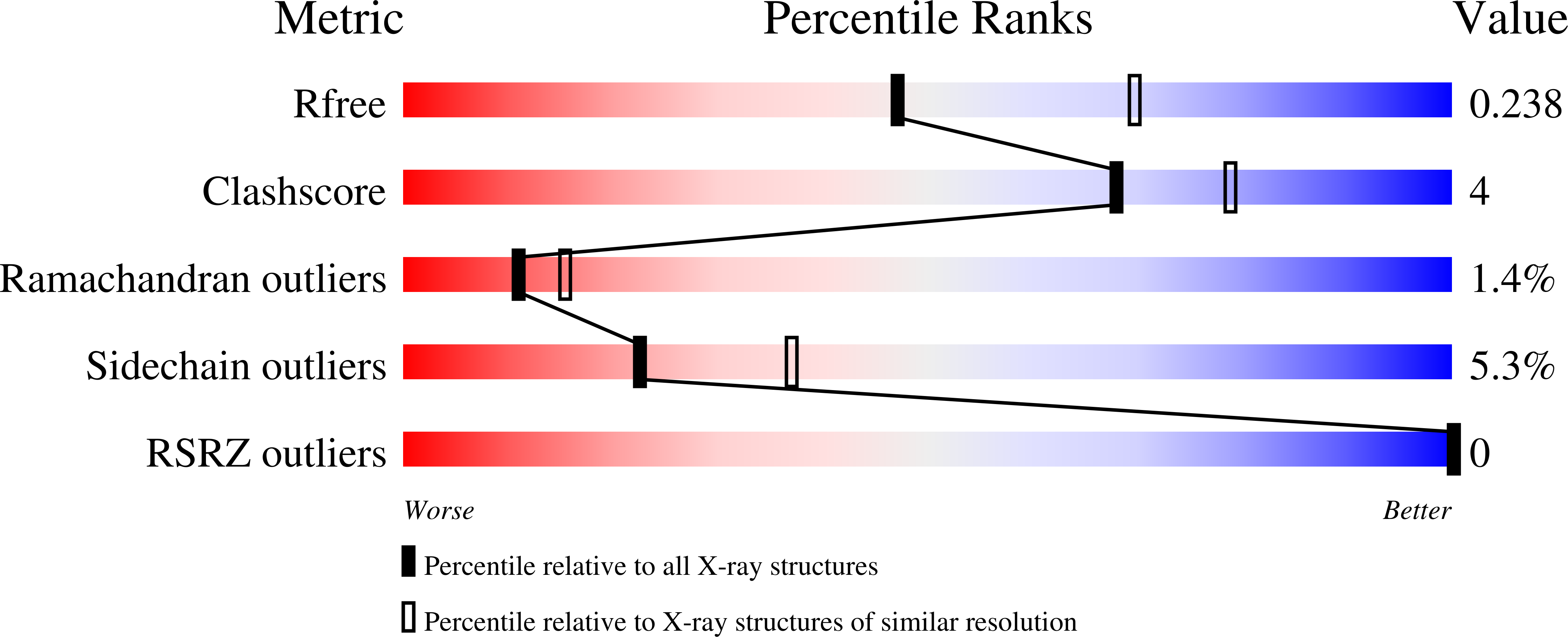
Resolution:
2.40 Å
R-Value Free:
0.26
R-Value Work:
0.22
R-Value Observed:
0.22
Space Group:
P 32