3VDY image
Deposition Date 2012-01-06
Release Date 2012-03-14
Last Version Date 2024-02-28
Entry Detail
PDB ID:
3VDY
Title:
B. subtilis SsbB/ssDNA
Biological Source:
Source Organism:
Bacillus subtilis (Taxon ID: 1423)
Synthetic DNA (Taxon ID: 32630)
Host Organism:
Method Details:
Experimental Method:
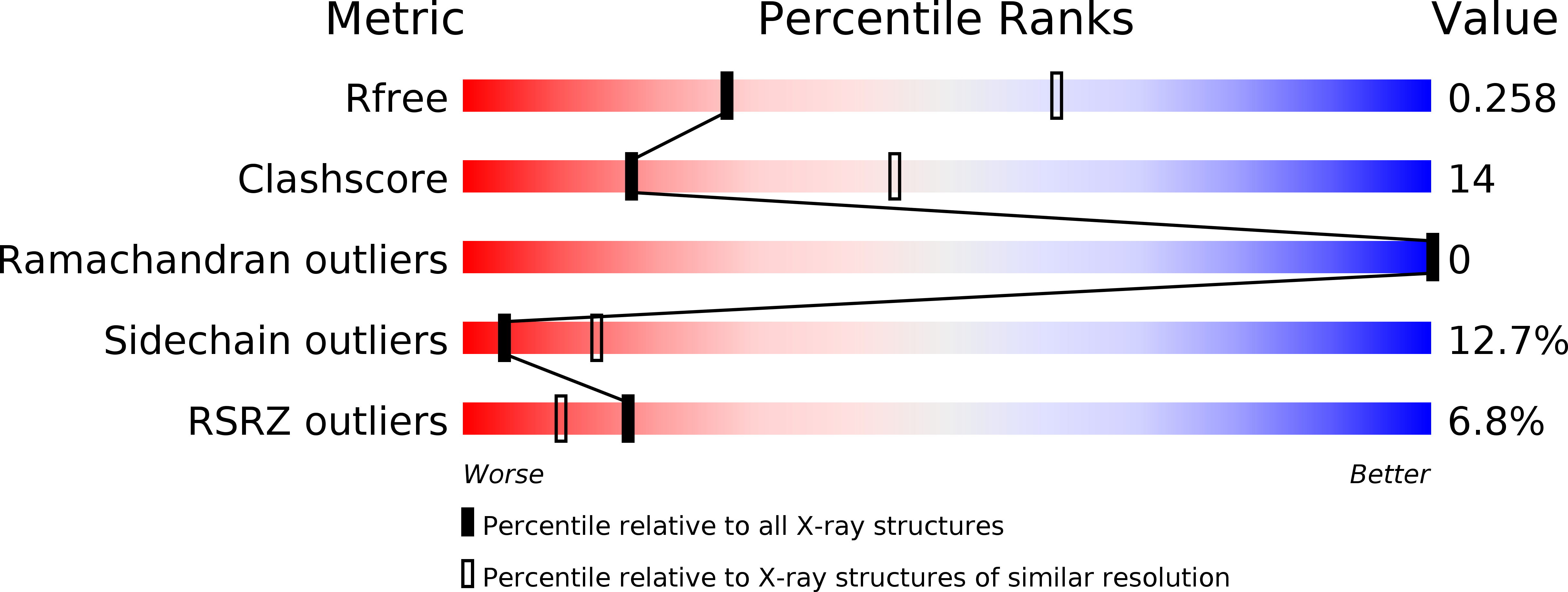
Resolution:
2.80 Å
R-Value Free:
0.26
R-Value Work:
0.22
R-Value Observed:
0.23
Space Group:
P 43 21 2
Macromolecular Entities
Structures with similar UniProt ID
Protein Blast
Polymer Type:polypeptide(L)
Molecule:Single-stranded DNA-binding protein ssbB
Gene (Uniprot):ssbB
Chain IDs:A, B
Chain Length:116
Number of Molecules:2
Biological Source:Bacillus subtilis
Polymer Type:polydeoxyribonucleotide
Molecule:5'-D(P*TP*TP*TP*TP*TP*TP*TP*TP*TP*TP*TP*TP*TP*TP*TP*TP*TP*TP*TP*TP*TP*TP*TP*TP*TP*TP*TP*TP*TP*TP*TP*TP*TP*TP*T)-3'
Chain IDs:C, D, E, F, G
Chain Length:35
Number of Molecules:5
Biological Source:Synthetic DNA
Ligand Molecules
Primary Citation
Genetic recombination in Bacillus subtilis: a division of labor between two single-strand DNA-binding proteins.
Nucleic Acids Res. 40 5546 5559 (2012)
PMID: 22373918 DOI: 10.1093/nar/gks173

Abstact

We have investigated the structural, biochemical and cellular roles of the two single-stranded (ss) DNA-binding proteins from Bacillus subtilis, SsbA and SsbB. During transformation, SsbB localizes at the DNA entry pole where it binds and protects internalized ssDNA. The 2.8-Å resolution structure of SsbB bound to ssDNA reveals a similar overall protein architecture and ssDNA-binding surface to that of Escherichia coli SSB. SsbA, which binds ssDNA with higher affinity than SsbB, co-assembles onto SsbB-coated ssDNA and the two proteins inhibit ssDNA binding by the recombinase RecA. During chromosomal transformation, the RecA mediators RecO and DprA provide RecA access to ssDNA. Interestingly, RecO interaction with ssDNA-bound SsbA helps to dislodge both SsbA and SsbB from the DNA more efficiently than if the DNA is coated only with SsbA. Once RecA is nucleated onto the ssDNA, RecA filament elongation displaces SsbA and SsbB and enables RecA-mediated DNA strand exchange. During plasmid transformation, RecO localizes to the entry pole and catalyzes annealing of SsbA- or SsbA/SsbB-coated complementary ssDNAs to form duplex DNA with ssDNA tails. Our results provide a mechanistic framework for rationalizing the coordinated events modulated by SsbA, SsbB and RecO that are crucial for RecA-dependent chromosomal transformation and RecA-independent plasmid transformation.

Legend

Protein

Chemical

Disease

Primary Citation of related structures