3VDL image
Entry Detail
PDB ID:
3VDL
Keywords:
Title:
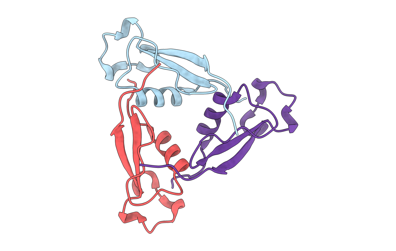
Crystal structure of circumsporozoite protein aTSR domain, P43212 form
Biological Source:
Source Organism:
Host Organism:
PDB Version:
Deposition Date:
2012-01-05
Release Date:
2012-05-09
Method Details:
Experimental Method:
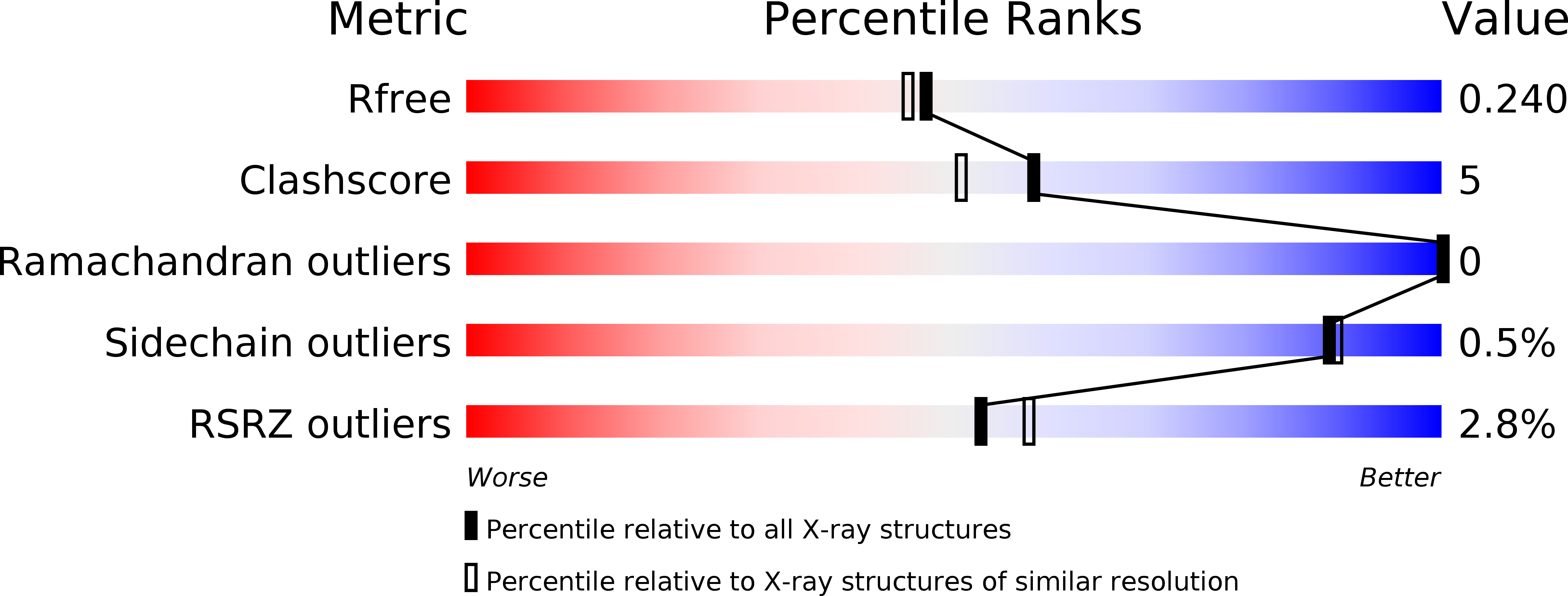
Resolution:
2.04 Å
R-Value Free:
0.24
R-Value Work:
0.19
R-Value Observed:
0.19
Space Group:
P 43 21 2
Macromolecular Entities
Polymer Type:polypeptide(L)
Description:Circumsporozoite (CS) protein
Chain IDs:A, B, C
Chain Length:77
Number of Molecules:3
Biological Source:Plasmodium falciparum
Primary Citation
Unexpected fold in the circumsporozoite protein target of malaria vaccines.
Proc.Natl.Acad.Sci.USA 109 7817 7822 (2012)
PMID: 22547819 DOI: 10.1073/pnas.1205737109

Abstact

Circumsporozoite (CS) protein is the major surface component of Plasmodium falciparum sporozoites and is essential for host cell invasion. A vaccine containing tandem repeats, region III, and thrombospondin type-I repeat (TSR) of CS is efficacious in phase III trials but gives only a 35% reduction in severe malaria in the first year postimmunization. We solved crystal structures showing that region III and TSR fold into a single unit, an "αTSR" domain. The αTSR domain possesses a hydrophobic pocket and core, missing in TSR domains. CS binds heparin, but αTSR does not. Interestingly, polymorphic T-cell epitopes map to specialized αTSR regions. The N and C termini are unexpectedly close, providing clues for sporozoite sheath organization. Elucidation of a unique structure of a domain within CS enables rational design of next-generation subunit vaccines and functional and medicinal chemical investigation of the conserved hydrophobic pocket.

Legend

Protein

Chemical

Disease

Primary Citation of related structures