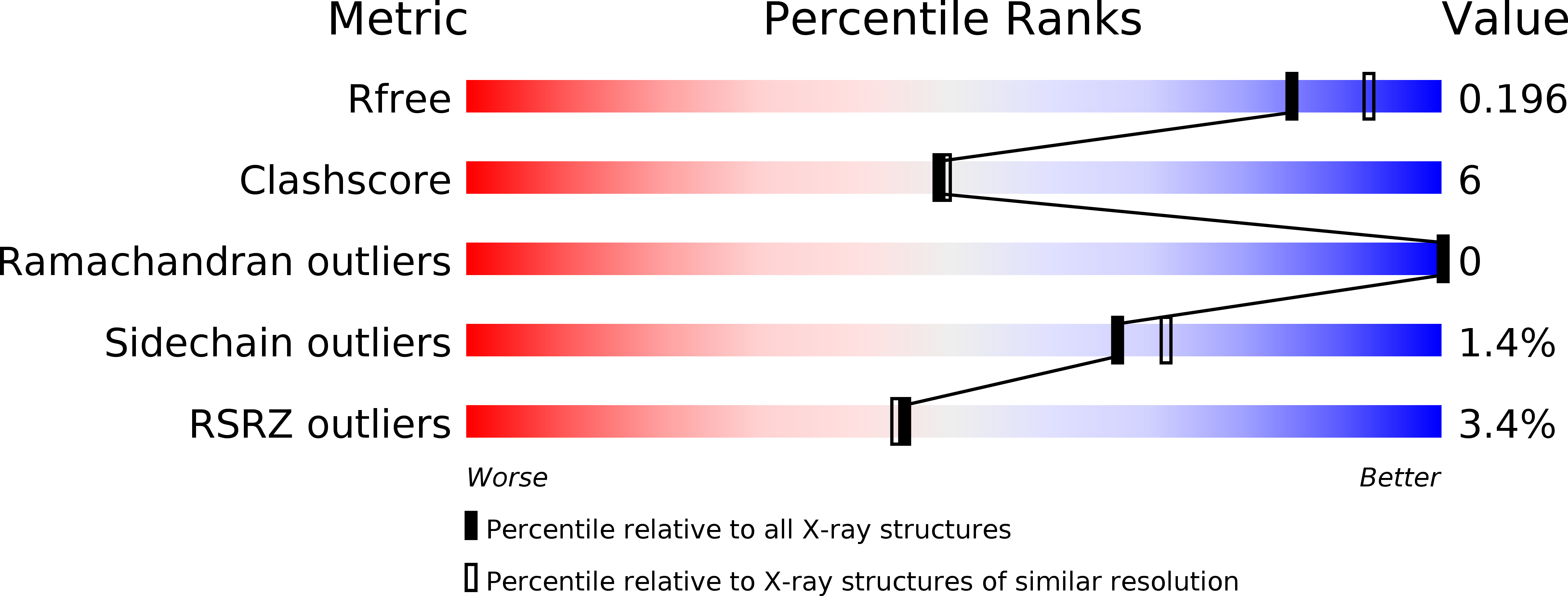
3VDI image
Entry Detail
PDB ID:
3VDI
Keywords:
Title:
Structure of the FMO protein from Pelodictyon phaeum
Biological Source:
Source Organism:
PDB Version:
Deposition Date:
2012-01-05
Release Date:
2012-01-25
Method Details:
Experimental Method:
Resolution:
1.99 Å
R-Value Free:
0.19
R-Value Work:
0.16
R-Value Observed:
0.16
Space Group:
P 63
Macromolecular Entities
Polymer Type:polypeptide(L)
Description:bacteriochlorophyll A protein
Chain IDs:A
Chain Length:367
Number of Molecules:1
Biological Source:Pelodictyon phaeum
Primary Citation
Reinterpretation of the electron density at the site of the eighth bacteriochlorophyll in the FMO protein from Pelodictyon phaeum.
Photosynth.Res. 112 71 74 (2012)
PMID: 22457093 DOI: 10.1007/s11120-012-9735-8

Abstact

The Fenna-Matthews-Olson antenna protein from the green bacterium Pelodictyon phaeum mediates the energy transfer from a peripheral antenna complex to the membrane-bound reaction center. The three-dimensional structure of this protein has been previously modeled using X-ray diffraction to a resolution limit of 2.0 Å, with R (work) and R (free) values of 16.6 and 19.9%, respectively (Larson et al., Photosynth Res 107:139-150, 2011). This model shows the protein as consisting of β-sheets surrounding several bacteriochlorophyll cofactors. While most of the model clearly matches the electron density maps, in this paper we re-examine the electron density for a specific feature, namely the eighth bacteriochlorophyll a cofactor. This electron density is now interpreted as arising primarily from the end of an otherwise disordered polyethylene glycol molecule. Additional electron density is present but the density is weak and cannot be unambiguously assigned. The new model has R (work) and R (free) values of 16.2 and 19.0%, respectively.

Legend

Protein

Chemical

Disease

Primary Citation of related structures