
Deposition Date
2012-01-04
Release Date
2012-04-18
Last Version Date
2024-10-30
Entry Detail
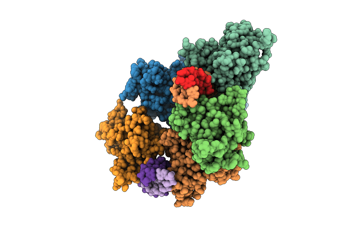
PDB ID:
3VD2
Keywords:
Title:
structure of p73 DNA binding domain tetramer modulates p73 transactivation
Biological Source:
Source Organism(s):
Homo sapiens (Taxon ID: 9606)
Expression System(s):
Method Details:
Experimental Method:
Resolution:
4.00 Å
R-Value Free:
0.28
R-Value Work:
0.24
R-Value Observed:
0.24
Space Group:
C 1 2 1
