Deposition Date
2012-01-03
Release Date
2012-01-11
Last Version Date
2024-10-30
Entry Detail
PDB ID:
3VC8
Keywords:
Title:
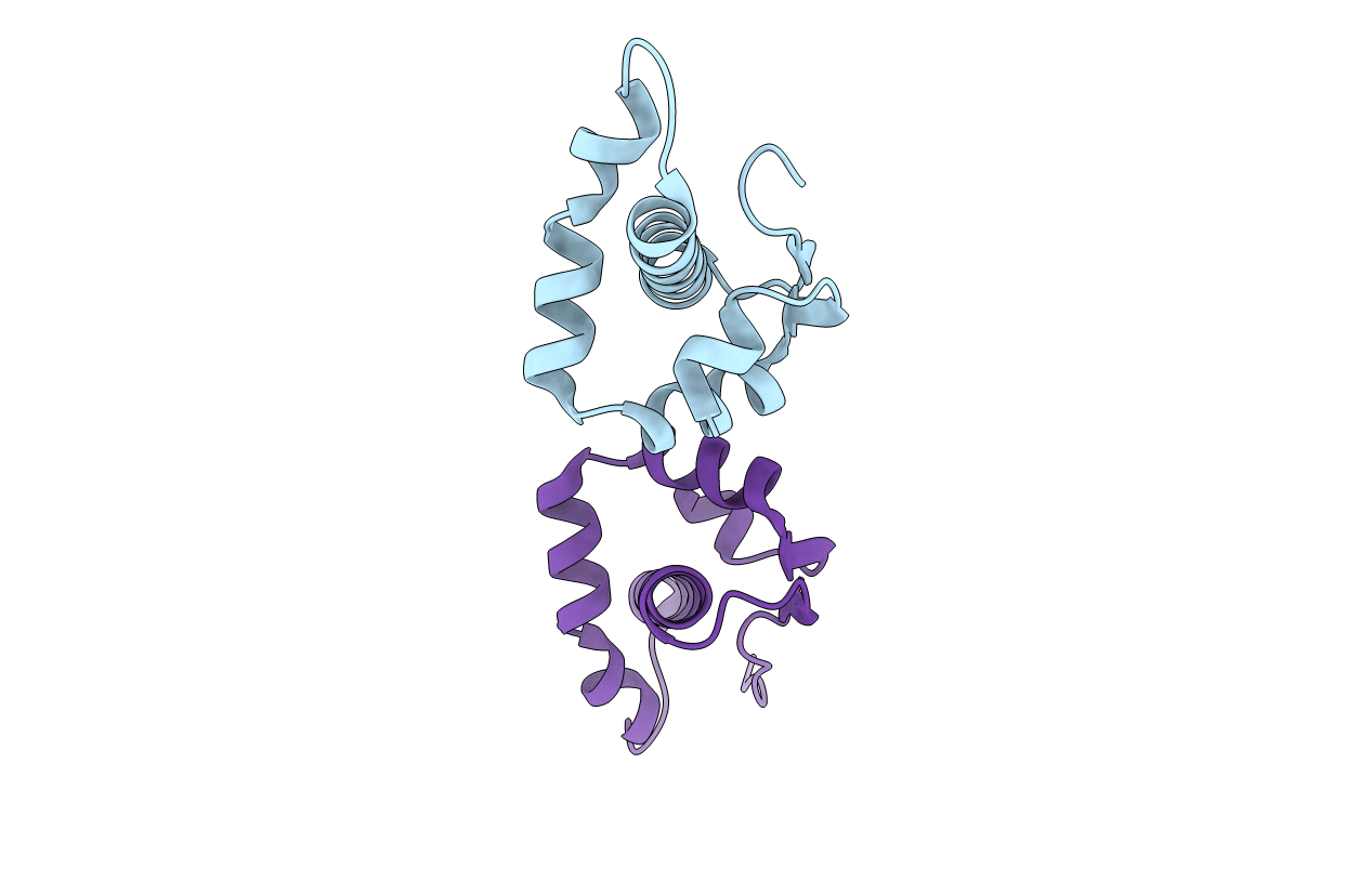
Crystal structure of the C-terminal cytoplasmic domain of non-structural protein 4 from mouse hepatitis virus A59
Biological Source:
Source Organism(s):
Murine hepatitis virus (Taxon ID: 591071)
Expression System(s):
Method Details:
Experimental Method:
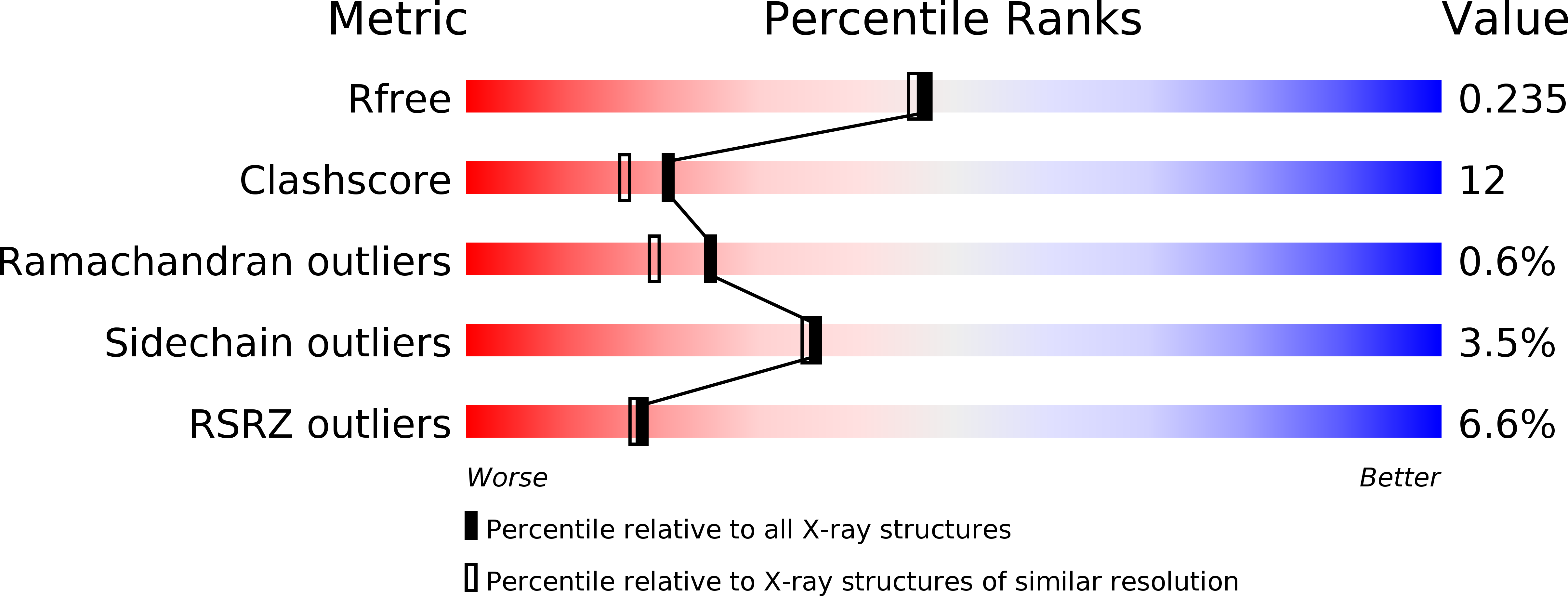
Resolution:
2.00 Å
R-Value Free:
0.26
R-Value Work:
0.22
Space Group:
C 1 2 1