3VBZ image
Deposition Date 2012-01-03
Release Date 2012-07-25
Last Version Date 2024-11-27
Entry Detail
PDB ID:
3VBZ
Keywords:
Title:
Crystal structure of Taipoxin beta subunit isoform 2
Biological Source:
Source Organism:
Method Details:
Experimental Method:
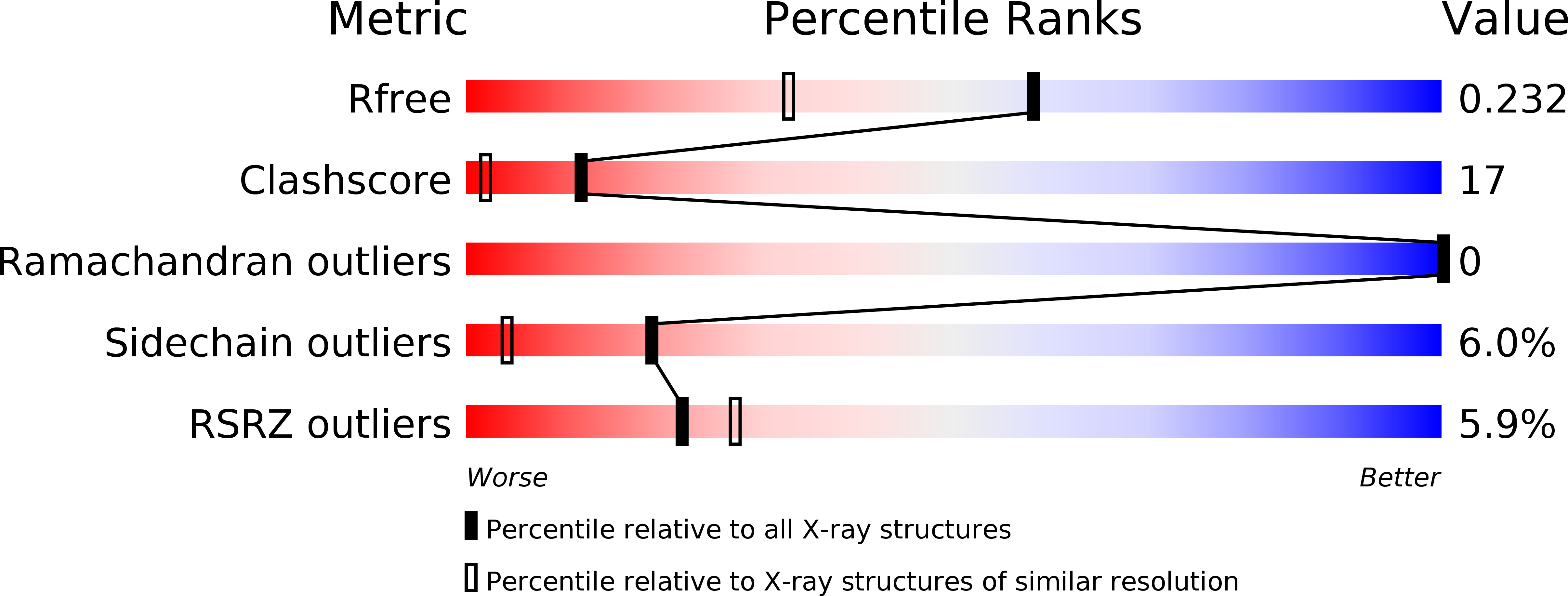
Resolution:
1.76 Å
R-Value Free:
0.22
R-Value Work:
0.18
R-Value Observed:
0.18
Space Group:
P 1 21 1
Macromolecular Entities
Polymer Type:polypeptide(L)
Molecule:Phospholipase A2 homolog, taipoxin beta chain
Chain IDs:A, B
Chain Length:118
Number of Molecules:2
Biological Source:Oxyuranus scutellatus scutellatus
Primary Citation
Structural analysis of trimeric phospholipase A(2) neurotoxin from the Australian taipan snake venom.
Febs J. 279 3121 3135 (2012)
PMID: 22776098 DOI: 10.1111/j.1742-4658.2012.08691.x

Abstact

Snake pre-synaptic neurotoxins endowed with phospholipase A(2) activity are potent inducers of paralysis through the specific disruption of the neuromuscular junction pre-synaptic membrane and represent a valuable tool for investigating neuronal degeneration and recovery. They have different structural complexity and a wide range of lethal potency and enzymatic activity, although they share a similar mechanism of action. Although no correlation has been reported between neurotoxicity and enzymatic activity, toxicity increases with structural complexity and phospholipase A(2) oligomers show 10-fold lower LD(50) values compared to their monomeric counterparts. To date, no structural study has been performed on multimeric SPANs with the aim of shedding light on the correlation between structural complexity and neurotoxicity. In the present study, we investigated the structure of taipoxin, a trimeric phospholipase A(2) neurotoxin, as well as that of its subunits, by X-ray crystallography and small angle X-ray scattering analysis. We present the high-resolution structure of two isoforms of the taipoxin β subunit, which show no neurotoxic activity but enhance the activity of the other subunits in the complex. One isoform shows no structural change that could justify the lack of activity. The other displays three point mutations in critical positions for the catalytic activity. Moreover, we designed a model for the quaternary structure of taipoxin under physiological conditions, in which the three subunits are organized into a flat holotoxin with the substrate binding sockets exposed on the same side of the complex, which suggests a role for this interface in the toxin-membrane interaction.

Legend

Protein

Chemical

Disease

Primary Citation of related structures