3VA2 image
Deposition Date 2011-12-28
Release Date 2012-07-25
Last Version Date 2024-11-20
Entry Detail
PDB ID:
3VA2
Title:
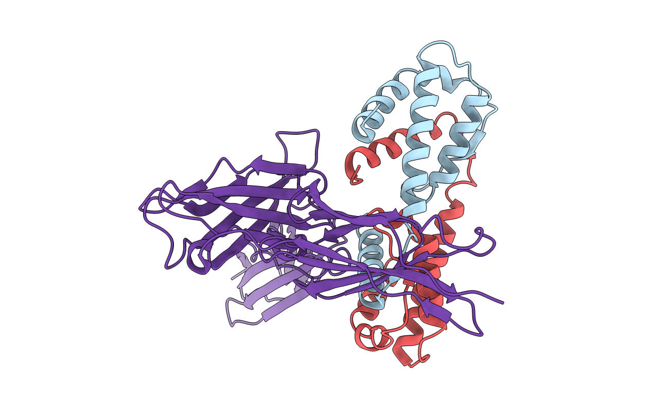
Crystal structure of human Interleukin-5 in complex with its alpha receptor
Biological Source:
Source Organism:
Homo sapiens (Taxon ID: 9606)
Host Organism:
Method Details:
Experimental Method:
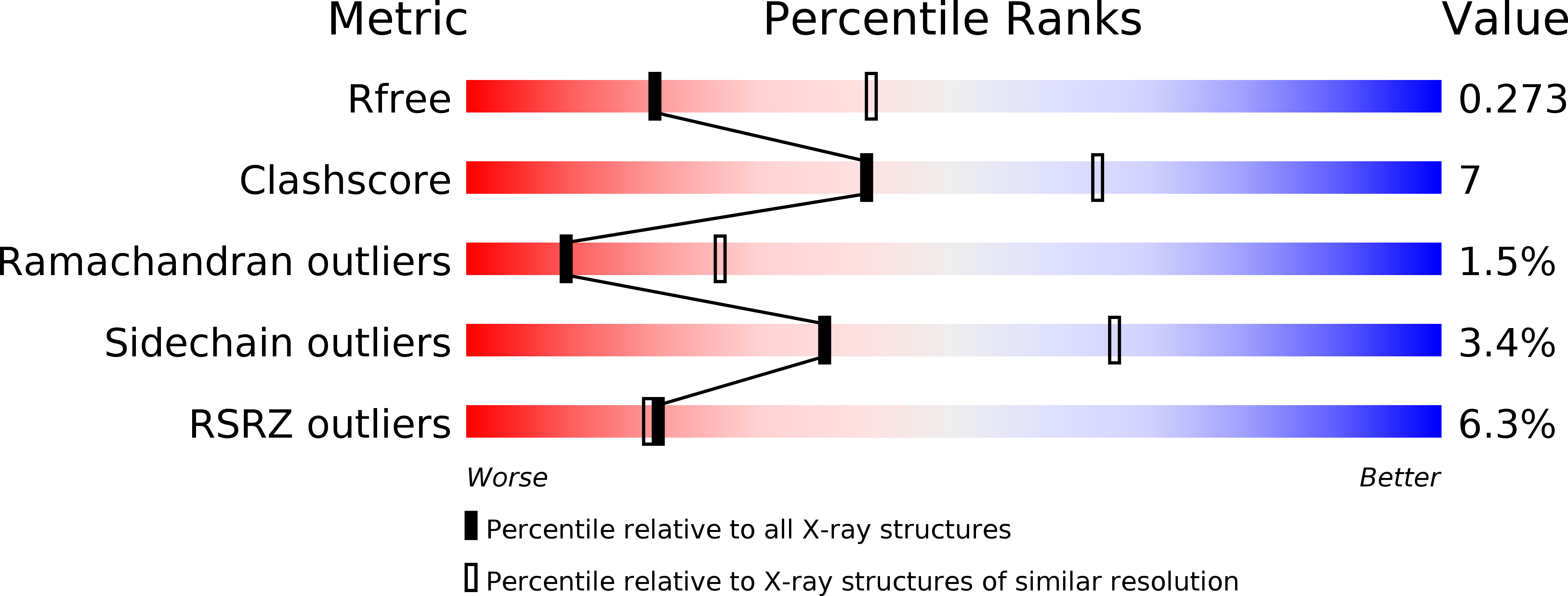
Resolution:
2.70 Å
R-Value Free:
0.27
R-Value Work:
0.22
R-Value Observed:
0.22
Space Group:
P 21 21 21
Macromolecular Entities
Structures with similar UniProt ID
Protein Blast
Polymer Type:polypeptide(L)
Molecule:Interleukin-5
Gene (Uniprot):IL5
Chain IDs:A, B
Chain Length:119
Number of Molecules:2
Biological Source:Homo sapiens
Structures with similar UniProt ID
Protein Blast
Polymer Type:polypeptide(L)
Molecule:Interleukin-5 receptor subunit alpha
Gene (Uniprot):IL5RA
Chain IDs:C
Chain Length:322
Number of Molecules:1
Biological Source:Homo sapiens
Primary Citation
Structural basis of interleukin-5 dimer recognition by its alpha receptor
Protein Sci. 21 850 864 (2012)
PMID: 22528658 DOI: 10.1002/pro.2072

Abstact

Interleukin-5 (IL-5), a major hematopoietin, stimulates eosinophil proliferation, migration, and activation, which have been implicated in the pathogenesis of allergic inflammatory diseases, such as asthma. The specific IL-5 receptor (IL-5R) consists of the IL-5 receptor α subunit (IL-5RA) and the common receptor β subunit (βc). IL-5 binding to IL-5R on target cells induces rapid tyrosine phosphorylation and activation of various cellular proteins, including JAK1/JAK2 and STAT1/STAT5. Here, we report the crystal structure of dimeric IL-5 in complex with the IL-5RA extracellular domains. The structure revealed that IL-5RA sandwiches the IL-5 homodimer by three tandem domains, arranged in a "wrench-like" architecture. This association mode was confirmed for human cells expressing IL-5 and the full-length IL-5RA by applying expanded genetic code technology: protein photo-cross-linking experiments revealed that the two proteins interact with each other in vivo in the same manner as that in the crystal structure. Furthermore, a comparison with the previously reported, partial GM-CSF•GM-CSFRA•βc structure enabled us to propose complete structural models for the IL-5 and GM-CSF receptor complexes, and to identify the residues conferring the cytokine-specificities of IL-5RA and GM-CSFRA.

Legend

Protein

Chemical

Disease

Primary Citation of related structures
Feedback Form
Name
Email
Institute
Feedback