Deposition Date
2011-12-27
Release Date
2013-01-23
Last Version Date
2024-11-06
Entry Detail
PDB ID:
3V9E
Keywords:
Title:
Structure of the L499M mutant of the laccase from B.aclada
Biological Source:
Source Organism(s):
Botrytis aclada (Taxon ID: 139639)
Expression System(s):
Method Details:
Experimental Method:
Resolution:
1.70 Å
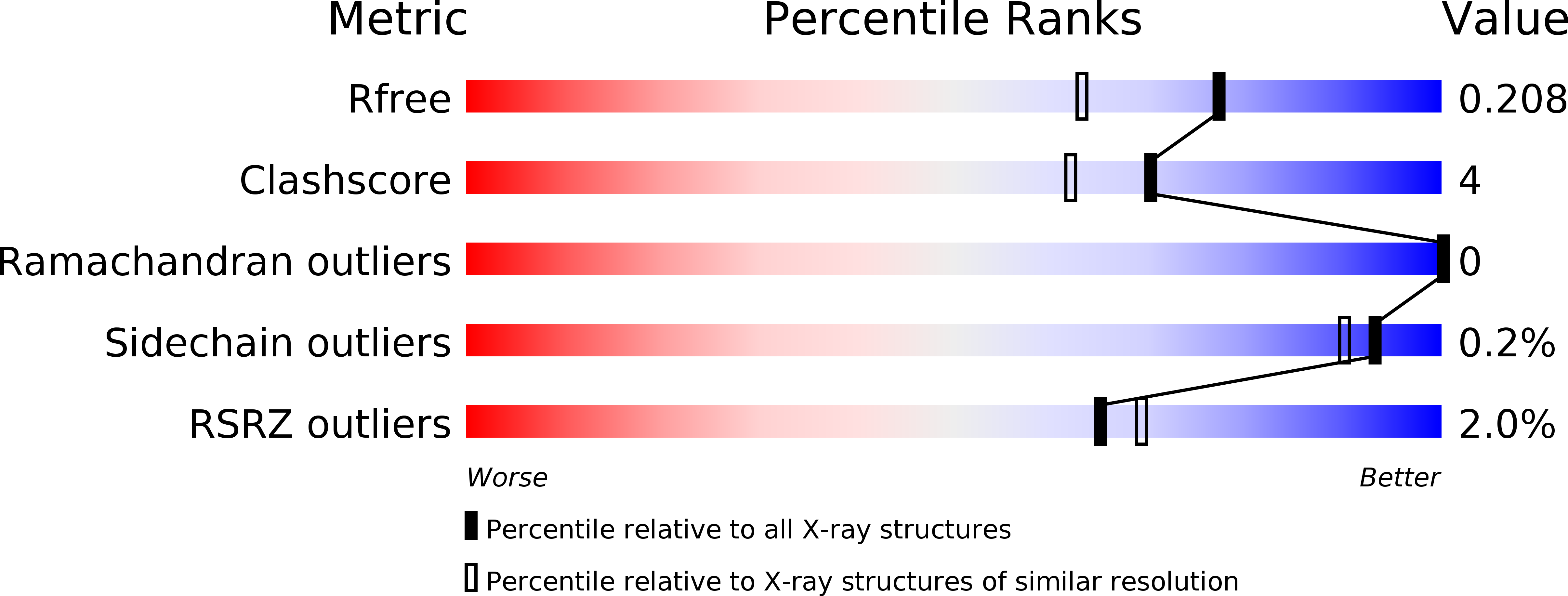
R-Value Free:
0.19
R-Value Work:
0.16
R-Value Observed:
0.16
Space Group:
C 1 2 1