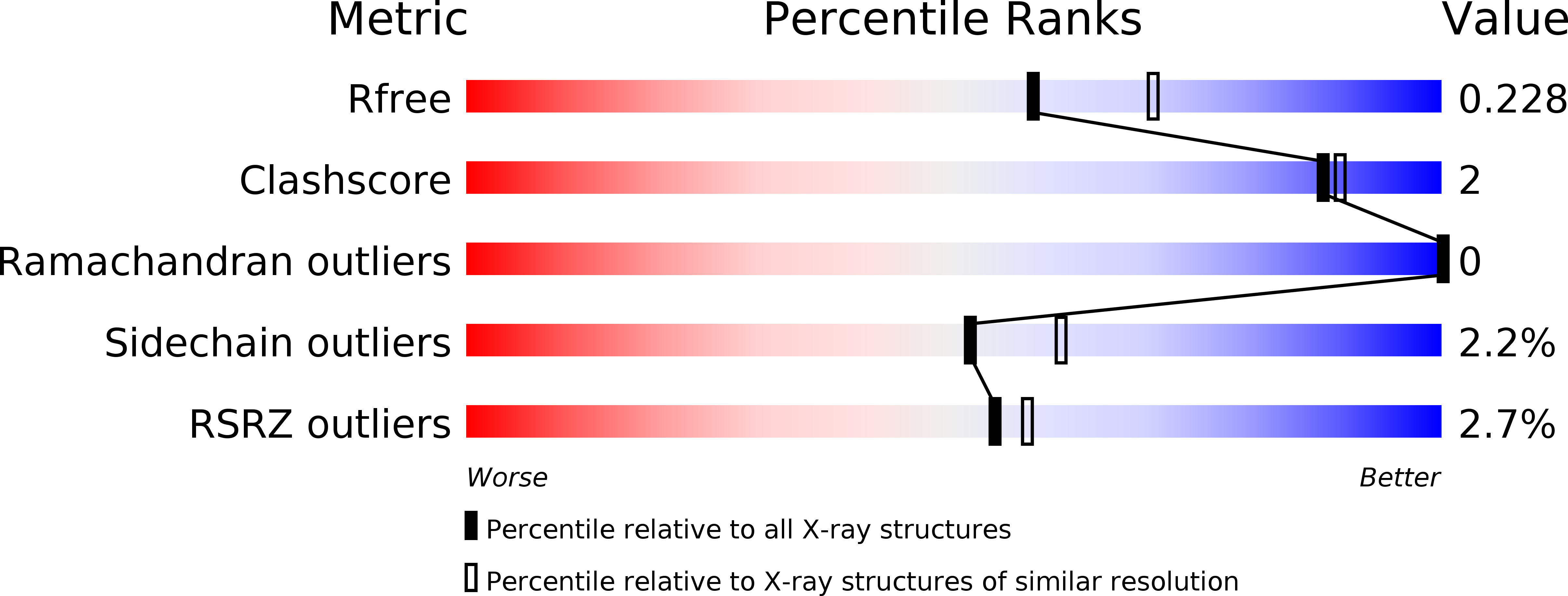
3V7O image
Deposition Date 2011-12-21
Release Date 2012-02-08
Last Version Date 2025-10-22
Entry Detail
PDB ID:
3V7O
Keywords:
Title:
Crystal structure of the C-terminal domain of Ebola virus VP30 (strain Reston-89)
Biological Source:
Source Organism:
Host Organism:
Method Details:
Experimental Method:
Resolution:
2.25 Å
R-Value Free:
0.22
R-Value Work:
0.19
R-Value Observed:
0.19
Space Group:
P 21 21 21
Macromolecular Entities
Structures with similar UniProt ID
Protein Blast
Polymer Type:polypeptide(L)
Molecule:Minor nucleoprotein VP30
Gene (Uniprot):VP30
Chain IDs:A, B
Chain Length:227
Number of Molecules:2
Biological Source:Reston ebolavirus
Ligand Molecules
Primary Citation
Structure of the Reston ebolavirus VP30 C-terminal domain.
Acta Crystallogr F Struct Biol Commun 70 457 460 (2014)
PMID: 24699737 DOI: 10.1107/S2053230X14003811

Abstact

The ebolaviruses can cause severe hemorrhagic fever. Essential to the ebolavirus life cycle is the protein VP30, which serves as a transcriptional cofactor. Here, the crystal structure of the C-terminal, NP-binding domain of VP30 from Reston ebolavirus is presented. Reston VP30 and Ebola VP30 both form homodimers, but the dimeric interfaces are rotated relative to each other, suggesting subtle inherent differences or flexibility in the dimeric interface.

Legend

Protein

Chemical

Disease

Primary Citation of related structures