Deposition Date
2011-12-20
Release Date
2012-02-08
Last Version Date
2024-11-27
Entry Detail
PDB ID:
3V7A
Keywords:
Title:
Structural basis for broad detection of genogroup II noroviruses by a monoclonal antibody that binds to a site occluded in the viral particle
Biological Source:
Source Organism(s):
Human calicivirus (Taxon ID: 150080)
Mus musculus (Taxon ID: 10090)
Mus musculus (Taxon ID: 10090)
Expression System(s):
Method Details:
Experimental Method:
Resolution:
3.30 Å
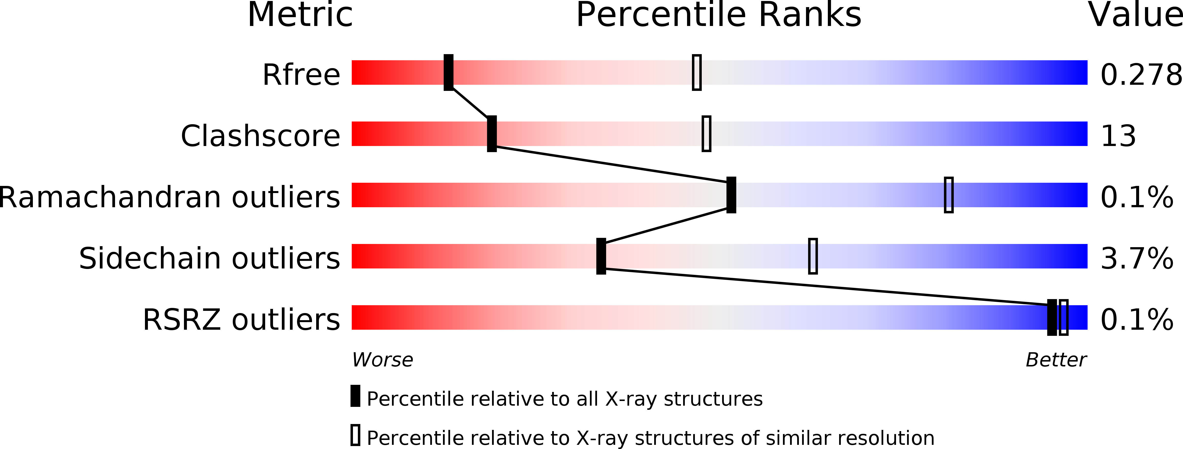
R-Value Free:
0.28
R-Value Work:
0.22
R-Value Observed:
0.22
Space Group:
P 43 2 2