3V6Q image
Deposition Date 2011-12-20
Release Date 2012-02-15
Last Version Date 2024-10-16
Entry Detail
PDB ID:
3V6Q
Keywords:
Title:
Crystal structure of the complex of bovine lactoperoxidase with Carbon monoxide at 2.0 A resolution
Biological Source:
Source Organism:
Bos taurus (Taxon ID: 9913)
Method Details:
Experimental Method:
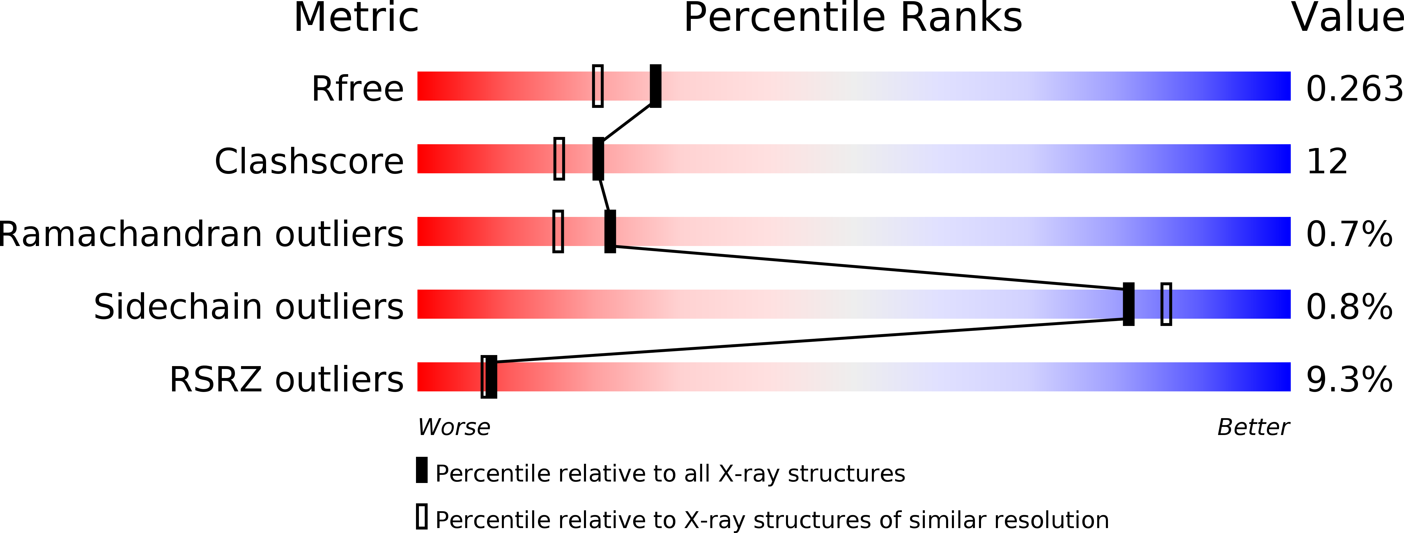
Resolution:
2.00 Å
R-Value Free:
0.24
R-Value Work:
0.19
R-Value Observed:
0.19
Space Group:
P 1 21 1
Macromolecular Entities
Polymer Type:polypeptide(L)
Molecule:Lactoperoxidase
Gene (Uniprot):LPO
Chain IDs:A
Chain Length:595
Number of Molecules:1
Biological Source:Bos taurus
Modified Residue
Compound ID Chain ID Parent Comp ID Details 2D Image
ASN A ASN GLYCOSYLATION SITE
SEP A SER PHOSPHOSERINE
Primary Citation
Bovine carbonyl lactoperoxidase structure at 2.0 angstrom resolution and infrared spectra as a function of pH.
Protein J. 31 598 608 (2012)
PMID: 22886082 DOI: 10.1007/s10930-012-9436-3

Abstact

Lactoperoxidase (LPO) is a hemeprotein catalyzing the oxidation of thiocyanate and I(-) into antimicrobials and small aromatic organics after being itself oxidized by H(2)O(2). LPO is excreted by the lungs, mammary glands, found in saliva and tears and protects mammals against bacterial, fungal and viral invasion. The Fe(II) form binds CO which inactivates LPO like many other hemeproteins. We present the 3-dimensional structure of CO-LPO at 2.0Å resolution and infrared (IR) spectra of the iron-bound CO stretch from pH 3 to 8.8 at 1 cm(-1) resolution. The observed Fe-C-O bond angle of 132° is more acute than the electronically related Fe(III), CN-LPO with a Fe-C-N angle of 161°. The orientations of the two ligands are different with the oxygen of CO pointing towards the imidazole of distal His109 while the nitrogen of CN points away, the Fe(II) moves towards His109 while the Fe(III) moves away; both movements are consistent with a hydrogen bond between the distal His109 and CO, but not to the nitrogen of CN-LPO. The IR spectra of CO-LPO exhibit two major CO absorbances with pH dependent relative intensities. Both crystallographic and IR data suggest proton donation to the CO oxygen by His109 with a pK ≈ 4; close to the pH of greatest enzyme turnover. The IR absorbance maxima are consistent with a first order correlation between frequency and Fe(III)/Fe(II) reduction potential at pH 7; both band widths at half-height correlate with electron density donation from Fe(II) to CO as gauged by the reduction potential.

Legend

Protein

Chemical

Disease

Primary Citation of related structures