3V6E image
Entry Detail
PDB ID:
3V6E
Title:
Crystal Structure of USP2 and a mutant form of Ubiquitin
Biological Source:
Source Organism:
Host Organism:
PDB Version:
Deposition Date:
2011-12-19
Release Date:
2012-12-19
Method Details:
Experimental Method:
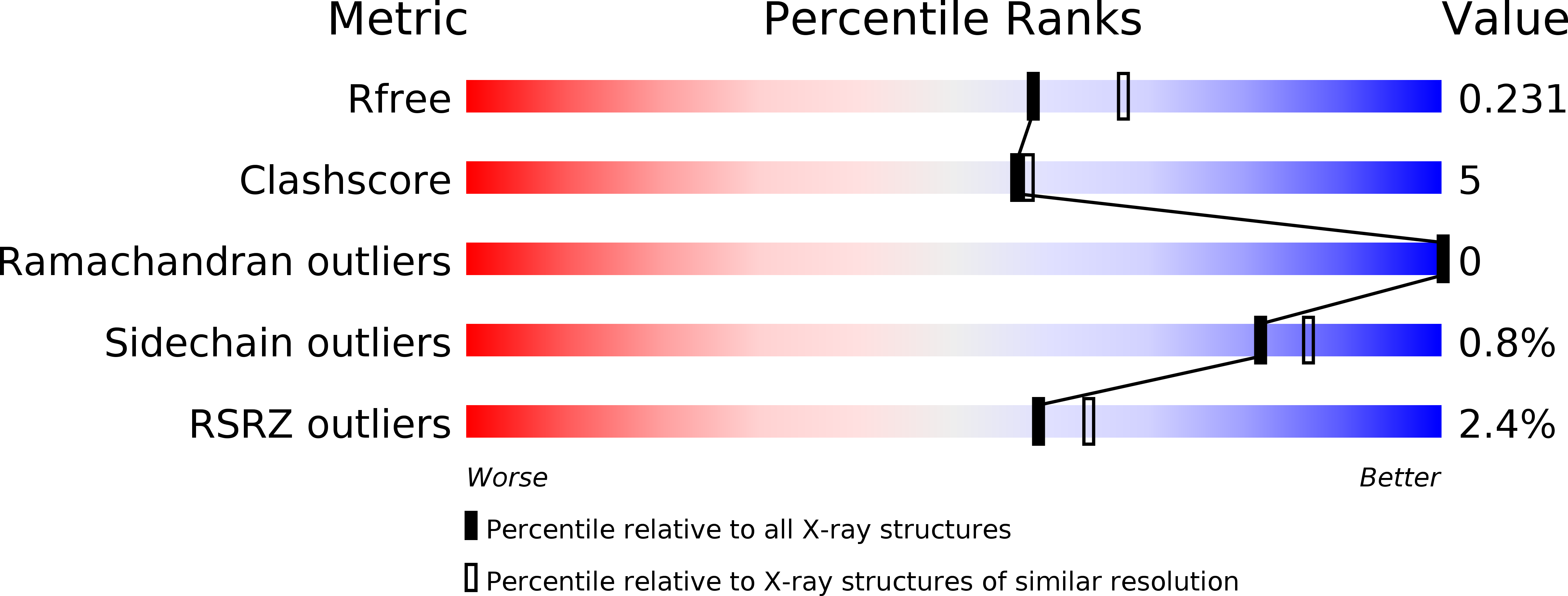
Resolution:
2.10 Å
R-Value Free:
0.23
R-Value Work:
0.17
R-Value Observed:
0.17
Space Group:
P 21 21 21
Macromolecular Entities
Polymer Type:polypeptide(L)
Description:Ubiquitin carboxyl-terminal hydrolase 2
Chain IDs:A
Chain Length:367
Number of Molecules:1
Biological Source:Homo sapiens
Polymer Type:polypeptide(L)
Description:Ubiquitin
Mutations:G76S, K82N, T85S, T88H
Chain IDs:B
Chain Length:91
Number of Molecules:1
Biological Source:Homo sapiens
Primary Citation

Abstact

The ubiquitin system regulates virtually all aspects of cellular function. We report a method to target the myriad enzymes that govern ubiquitination of protein substrates. We used massively diverse combinatorial libraries of ubiquitin variants to develop inhibitors of four deubiquitinases (DUBs) and analyzed the DUB-inhibitor complexes with crystallography. We extended the selection strategy to the ubiquitin conjugating (E2) and ubiquitin ligase (E3) enzymes and found that ubiquitin variants can also enhance enzyme activity. Last, we showed that ubiquitin variants can bind selectively to ubiquitin-binding domains. Ubiquitin variants exhibit selective function in cells and thus enable orthogonal modulation of specific enzymatic steps in the ubiquitin system.

Legend

Protein

Chemical

Disease

Primary Citation of related structures