Deposition Date
2011-12-16
Release Date
2012-10-03
Last Version Date
2023-09-13
Entry Detail
PDB ID:
3V57
Keywords:
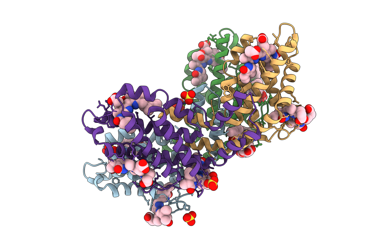
Title:
Crystal Structure of the B-phycoerythrin from the red algae Porphyridium Cruentum at pH8
Biological Source:
Source Organism(s):
Porphyridium purpureum (Taxon ID: 35688)
Method Details:
Experimental Method:
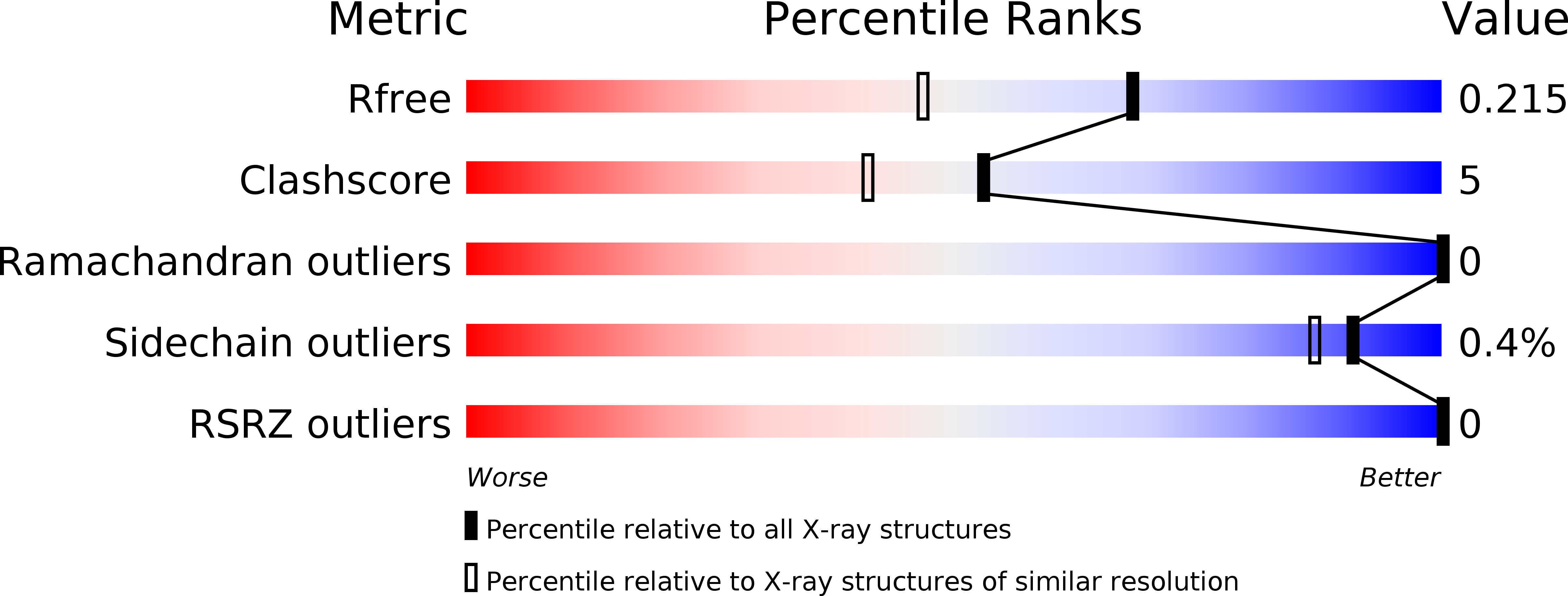
Resolution:
1.70 Å
R-Value Free:
0.21
R-Value Work:
0.17
R-Value Observed:
0.17
Space Group:
H 3