3V4R image
Entry Detail
PDB ID:
3V4R
Keywords:
Title:
Crystal structure of a UvrB dimer-DNA complex
Biological Source:
Source Organism:
Host Organism:
PDB Version:
Deposition Date:
2011-12-15
Release Date:
2012-07-04
Method Details:
Experimental Method:
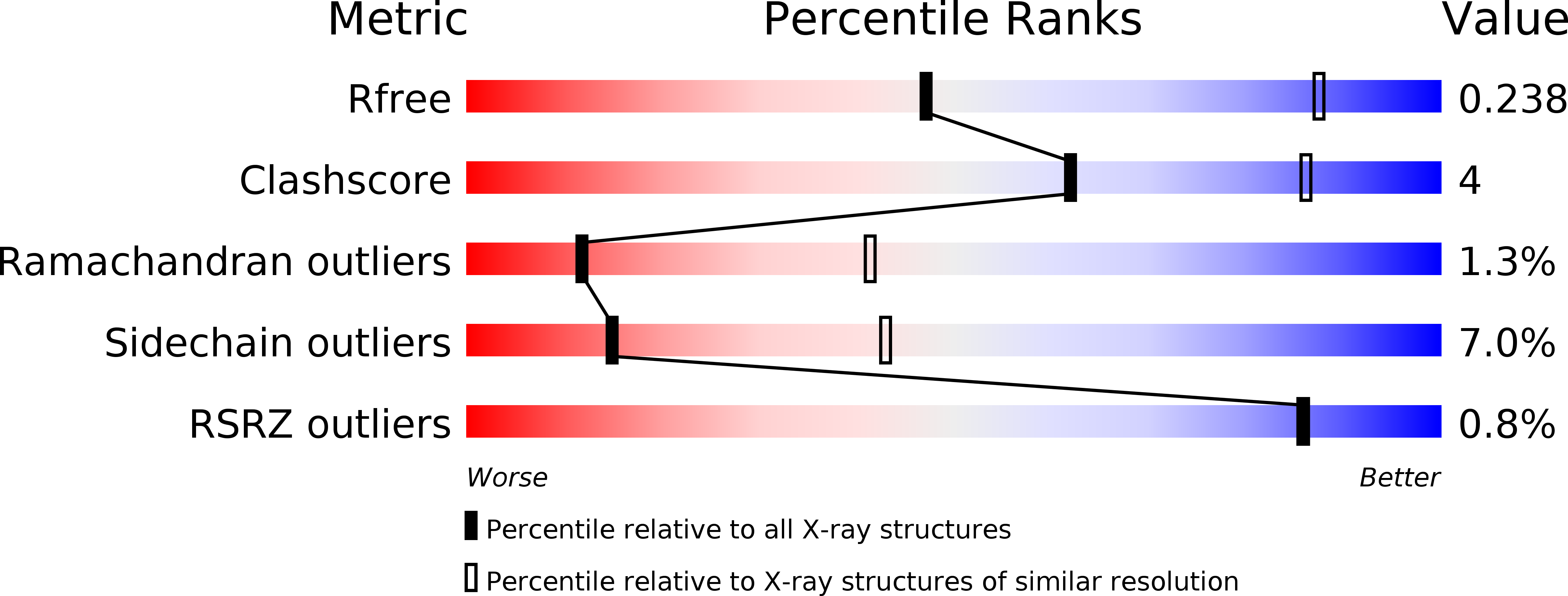
Resolution:
3.25 Å
R-Value Free:
0.21
R-Value Work:
0.17
R-Value Observed:
0.18
Space Group:
P 21 21 21
Macromolecular Entities
Polymer Type:polypeptide(L)
Description:UvrABC system protein B
Chain IDs:A, B
Chain Length:667
Number of Molecules:2
Biological Source:Bacillus subtilis
Polymer Type:polydeoxyribonucleotide
Description:DNA: 5 -TACTGTTT-3
Chain IDs:C, D
Chain Length:8
Number of Molecules:2
Biological Source:
Ligand Molecules
Primary Citation
Crystal structure of the UvrB dimer: insights into the nature and functioning of the UvrAB damage engagement and UvrB-DNA complexes.
Nucleic Acids Res. 40 8743 8758 (2012)
PMID: 22753105 DOI: 10.1093/nar/gks633

Abstact

UvrB has a central role in the highly conserved UvrABC pathway functioning not only as a damage recognition element but also as an essential component of the lesion tracking machinery. While it has been recently confirmed that the tracking assembly comprises a UvrA2B2 heterotetramer, the configurations of the damage engagement and UvrB-DNA handover complexes remain obscure. Here, we present the first crystal structure of a UvrB dimer whose biological significance has been verified using both chemical cross-linking and electron paramagnetic resonance spectroscopy. We demonstrate that this dimeric species stably associates with UvrA and forms a UvrA2B2-DNA complex. Our studies also illustrate how signals are transduced between the ATP and DNA binding sites to generate the helicase activity pivotal to handover and formation of the UvrB2-DNA complex, providing key insights into the configurations of these important repair intermediates.

Legend

Protein

Chemical

Disease

Primary Citation of related structures