3V4N image
Deposition Date 2011-12-15
Release Date 2012-04-25
Last Version Date 2024-10-16
Entry Detail
PDB ID:
3V4N
Title:
The Biochemical and Structural Basis for Inhibition of Enterococcus faecalis HMG-CoA Synthatse, mvaS, by Hymeglusin
Biological Source:
Source Organism(s):
Expression System(s):
Method Details:
Experimental Method:
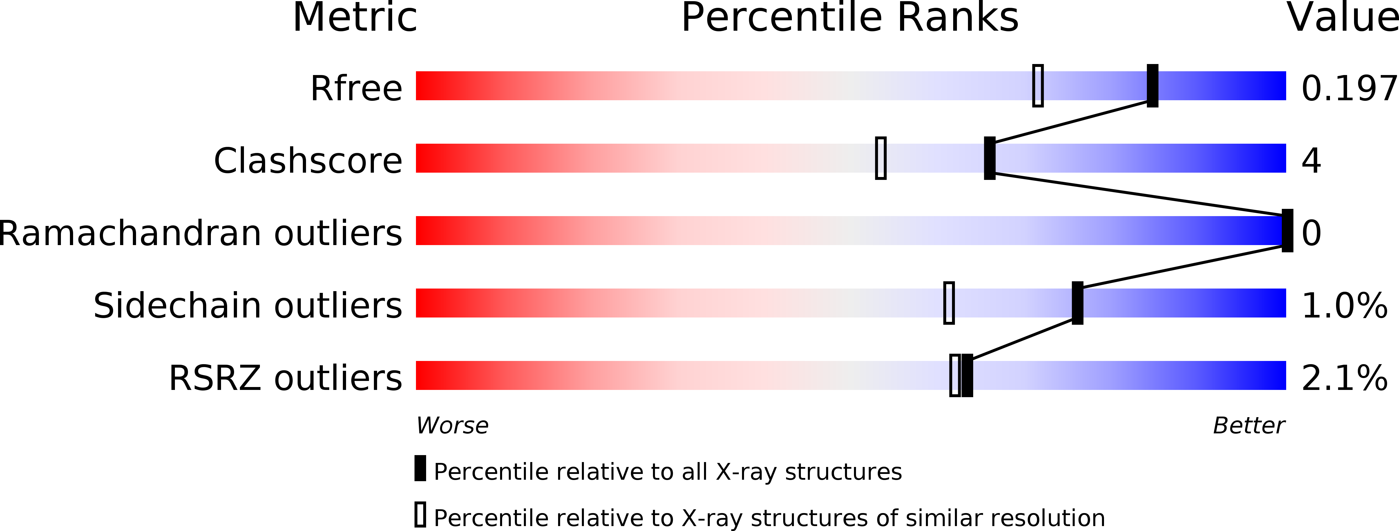
Resolution:
1.60 Å
R-Value Free:
0.19
R-Value Work:
0.14
R-Value Observed:
0.14
Space Group:
P 1 21 1
Macromolecular Entities
Structures with similar UniProt ID
Protein Blast
Polymer Type:polypeptide(L)
Molecule:HMG-CoA synthase
Gene (Uniprot):mvaS
Mutagens:A163S
Chain IDs:A, B, C, D
Chain Length:388
Number of Molecules:4
Biological Source:Enterococcus faecalis
Modified Residue
Compound ID Chain ID Parent Comp ID Details 2D Image
SNC A CYS S-NITROSO-CYSTEINE
Ligand Molecules
Primary Citation
Biochemical and structural basis for inhibition of Enterococcus faecalis hydroxymethylglutaryl-CoA synthase, mvaS, by hymeglusin.
Biochemistry 51 4713 4722 (2012)
PMID: 22510038 DOI: 10.1021/bi300037k

Abstact

Hymeglusin (1233A, F244, L-659-699) is established as a specific β-lactone inhibitor of eukaryotic hydroxymethylglutaryl-CoA synthase (HMGCS). Inhibition results from formation of a thioester adduct to the active site cysteine. In contrast, the effects of hymeglusin on bacterial HMG-CoA synthase, mvaS, have been minimally characterized. Hymeglusin blocks growth of Enterococcus faecalis. After removal of the inhibitor from culture media, a growth curve inflection point at 3.1 h is observed (vs 0.7 h for the uninhibited control). Upon hymeglusin inactivation of purified E. faecalis mvaS, the thioester adduct is more stable than that measured for human HMGCS. Hydroxylamine cleaves the thioester adduct; substantial enzyme activity is restored at a rate that is 8-fold faster for human HMGCS than for mvaS. Structural results explain these differences in enzyme-inhibitor thioester adduct stability and solvent accessibility. The E. faecalis mvaS-hymeglusin cocrystal structure (1.95 Å) reveals virtually complete occlusion of the bound inhibitor in a narrow tunnel that is largely sequestered from bulk solvent. In contrast, eukaryotic (Brassica juncea) HMGCS binds hymeglusin in a more solvent-exposed cavity.

Legend

Protein

Chemical

Disease

Primary Citation of related structures
Feedback Form
Name
Email
Institute
Feedback