Deposition Date
2011-12-14
Release Date
2012-01-04
Last Version Date
2024-10-09
Entry Detail
PDB ID:
3V4D
Keywords:
Title:
Crystal structure of RutC protein a member of the YjgF family from E.coli
Biological Source:
Source Organism(s):
Escherichia coli O6 (Taxon ID: 217992)
Expression System(s):
Method Details:
Experimental Method:
Resolution:
1.95 Å
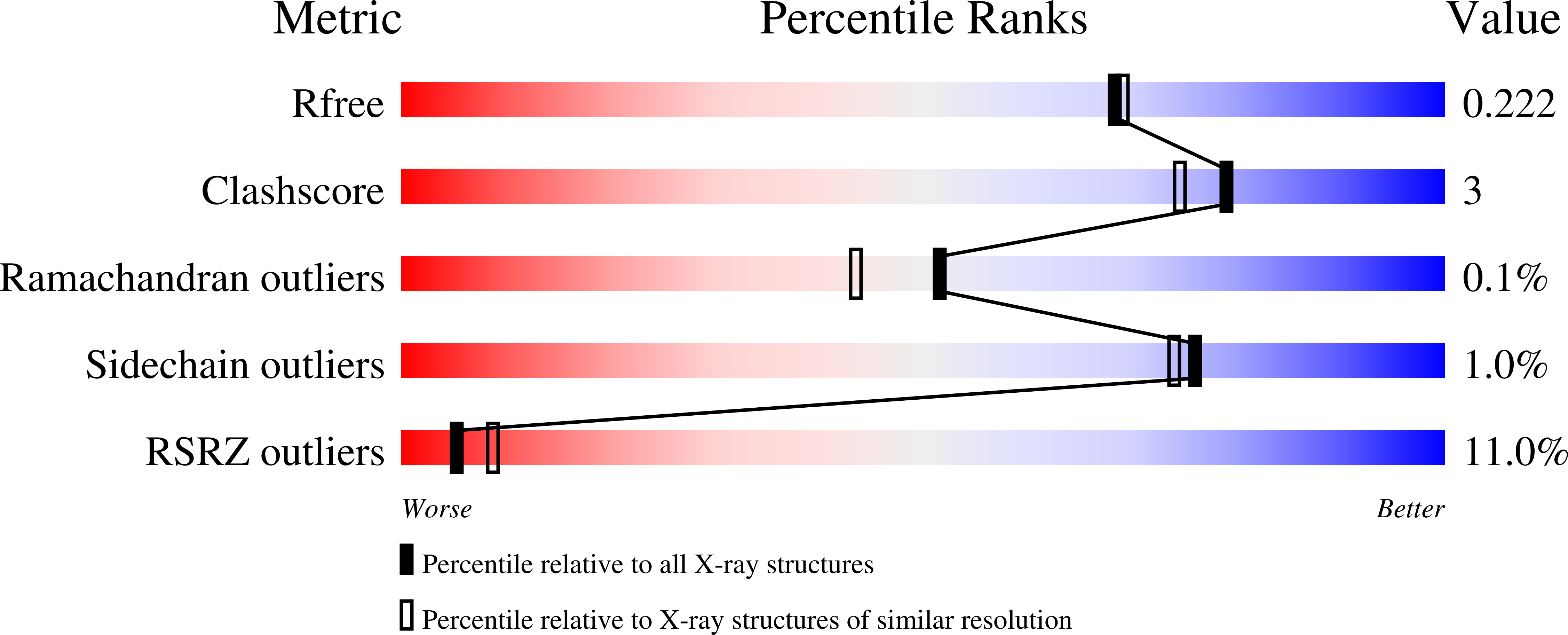
R-Value Free:
0.21
R-Value Work:
0.19
R-Value Observed:
0.19
Space Group:
P 21 21 2